Biomedical Engineering Reference
In-Depth Information
Fig. 8.7
PSF of a coherent microscope (positive real part shown)
Fig. 8.8
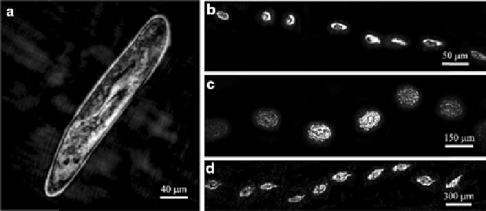
Images taken with underwater DHM. (
a
) Paramecium. (
b
-
d
) Trajectories of various
species swimming through the observation window, with a frame rate of 10 frames/s. (
b
) Ciliate;
(
c
), Didinium; (
d
) Rotifer (Reproduced with permission)
The scanning system is connected to the interferometer or OCT engine unit by a
fiber optic. Early time domain ophthalmic OCT systems (TD-OCT) used the
Michelson interferometer configuration as shown in Fig.
8.10
. In this case the
interference between object and reference paths is recorded as the depth scan is
performed by a mechanically driven mirror. OCT requires a bright light source of
limited temporal coherence and super-luminescent diodes, ultrashort pulse and
super-continuum lasers have all been used for this purpose [
18
].

Search WWH ::

Custom Search