Biomedical Engineering Reference
In-Depth Information
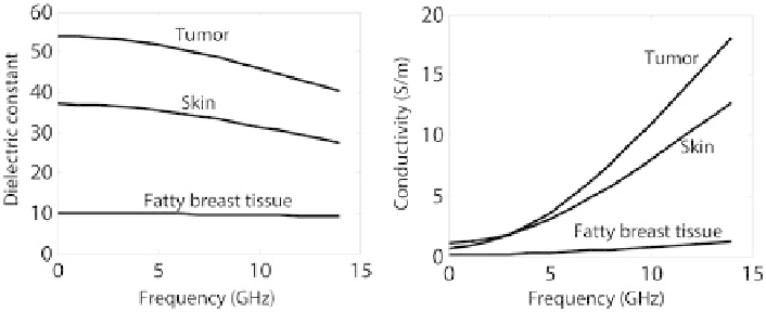
Fig. 1
Frequency dependence of the dielectric constant and conductivity for fatty breast tissue,
skin, and tumor. [
7
]
The main motivation for using ultra-narrow pulses for breast imaging arises from
the high contrast between the electrical properties of malignant and normal breast
tissues. For instance, this contrast is 5:1 for relative dielectric constant and 10:1
for conductivity at microwave frequencies [
3
,
4
] (see Fig.
1
). In addition to contrast,
UWB microwave imaging systems are also able to provide both adequate penetration
depth and necessary imaging resolution [
5
,
6
].
Medical Imaging for Breast Cancer
Among various types of medical imaging applications of UWB systems, breast cancer
detection is one of the most popular fields that has attracted much research interest.
In recent years, an increasing number of deaths due to breast cancer are reported.
Only in the USA, there is an average of 40,000 deaths a year because of this type
of cancer [
8
]. Since curing this type of cancer or long-term survival of patients are
highly dependent on early detection and timely medical intervention, a very precise
and dependable imaging system is required in the treatment procedure.
Conventional mammography has been performed using x-ray imaging of a com-
pressed breast for nonpalpable early stage breast cancer [
9
]. Throughout the time,
some technical advancements, such as digital mammography plus radiological ex-
pertise, have provided significant improvement of image quality and diagnosis using
x-ray. However, due to sensitivity issues of x-ray imaging, there are still many prob-
lems with relatively high false-negative rate of detection, and low positive predictive
rates resulting in many additional unnecessary biopsies [
10
]. In addition, in x-ray
imaging, patients have to deal with uncomfortable or painful breast compression and
exposure to low level ionization.
Usually, magnetic resonance imaging (MRI) and ultrasound are used for verifi-
cation of mammography detected lesions by x-ray. In these systems, the modalities
are not yet sensitive/specific enough or are too operator dependent or too costly to
be useful for screening purposes.
Search WWH ::

Custom Search