Biomedical Engineering Reference
In-Depth Information
Although most nonhuman primates tolerate bronchos-
copy well, a brief period of observation is required after the
procedure. The primate is assessed for respiratory difficulty
(stridor and dyspnea resulting from laryngeal edema, lar-
yngospasm, or bronchospasm) and monitored closely until
the effects of anesthetic wear off and gag reflex has
returned. If the primate has had a transbronchial biopsy,
a thoracic radiograph may be taken after the procedure to
rule out pneumothorax. Adverse affects of bronchoscopy
are infrequent. However, trauma to the airways, damage to
the vocal cords, excessive bleeding following biopsy, and
pneumothorax following lung biopsy can occur. Lar-
yngospasm is a rare complication but may sometimes
occur.
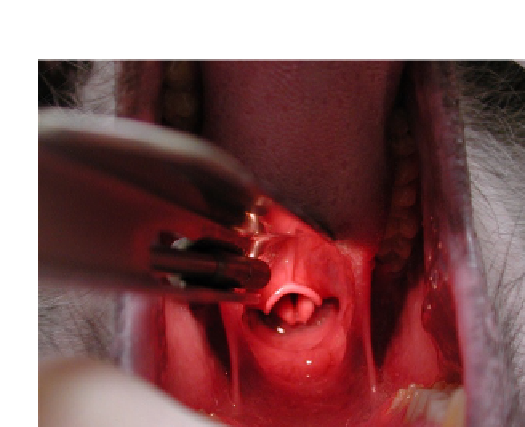
FIGURE 13.6
Oral cavity and pharynx of a nonhuman primate.
Ventral is up in this picture.
Pharyngeal Swabs
Pharyngeal swabs are used to collect specimens for
microbial cultures. For smaller nonhuman primates this
may be done without the aid of anesthesia but for larger
primates the use of anesthesia is advised. The area near the
crypts of the tonsils is usually the best area to obtain swabs
that have a greater percentage of positive bacterial cultures.
It is very important to quickly transfer the swab to a proper
culture media. This prevents the swab from quickly drying
out resulting in a false negative.
regurgitated, or for other complicating factors leading to
the threat of particles remaining in the mouth, the balloon
can be left slightly inflated to help remove any debris which
may remain in the trachea and pharynx.
Bronchoscopy
Bronchoscopy is a technique to visualize the inside of the
airways for diagnostic and therapeutic purposes. A bron-
choscope is inserted into the airways, usually through the
nose or mouth or occasionally through a tracheostomy.
Flexible bronchoscopy causes less discomfort for the
primate than rigid bronchoscopy and the procedure can
typically be performed easily and safely under light anes-
thesia. A pediatric endoscope of 3.8 mm outer diameter was
used for examination of rhesus monkeys (
Tate et al., 2004;
Singletary et al., 2008
) but may only pass to the level of the
main stem bronchi.
The nonhuman primate is administered an anesthetic
and atropine. Local anesthetics such as Marcaine 0.25%
mixed with sterile lube are often given to anesthetize the
mucous membranes of the pharynx, larynx, and trachea.
The flexible bronchoscope is inserted with the primate in
a sitting or supine position. If the subject is intubated, the
bronchoscope is inserted through an adapter connected to
the tracheal tube. The instrument is advanced to the trachea
and further down into the bronchial system and each area is
inspected as the bronchoscope passes. If an abnormality is
discovered, it may be biopsied, using a brush, a needle, or
forceps. A transbronchial lung biopsy may be obtained
using fluoroscopy.
Rigid bronchoscopy is performed under general anes-
thesia. Rigid bronchoscopes are too large to allow parallel
placement of other devices in the trachea; therefore the
primate is ventilated through the bronchoscope.
Tracheobronchial Washings
Tracheobronchial washings can be performed on
nonhuman primates that are anesthetized. Surgical expo-
sure of the trachea may be required for small nonhuman
primates with relatively flexible tracheas that lie deep
within the cervical musculature. The animal is sedated and
placed in dorsal recumbency and the skin directly above the
trachea is prepared aseptically. A surgical incision is made
on the ventral midline of the neck and the cervical muscles
are retracted laterally to provide exposure of the trachea.
The trachea may be elevated superficially using a hemostat
along its dorsal aspect. A catheter or needle is inserted
directly into the trachea and the flush and collection can
then be done. This technique cannot be repeated with any
frequency.
Another technique has also been described for
tracheobronchial lavage performed using an oral endotra-
cheal tube (
Ilievski and Fleischman, 1981
). Anesthetized
rhesus monkeys were placed in dorsal recumbency, with
their heads positioned in complete extension over the
table's edge. A 3 French, 40.6-cm feeding tube was used as
the flush catheter. The outer tube that the catheter was
supplied in was modified to serve as an endotracheal tube
and to protect the flush catheter from contamination by
oropharyngeal flora. A sterile straight hemostat or laryn-
goscope may be used for stabilization of the epiglottis.