Biomedical Engineering Reference
In-Depth Information
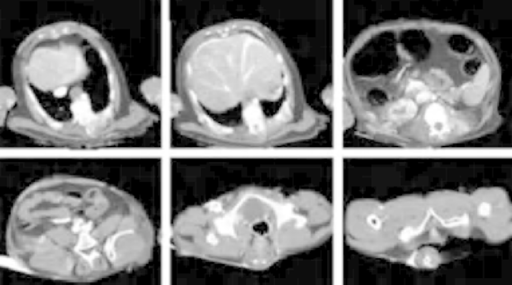
FIGURE 4.12
Axial CT images of abdomen, pelvis, and perineum (Papio anubis, adult female). (A) Section through caudal thorax and cranial
abdomen, showing the azygous lobe of the right lung positioned dorsal to the caudal (inferior) vena cava. The esophagus appears ventral to the vertebral
column and the descending aorta is to its immediate left. (B) Section through cranial abdomen illustrating the extent of the liver into the left as well as right
cranial quadrants of the abdominal cavity. (C) Abdominal cavity at the left of the pancreas and splenic hilum. Also visible is the right kidney, although the
left is not visible in this section as it is situated in a more caudal plane. The ascending colon is apparent ventral to the right kidney, as is the descending
colon ventral to the spleen. (D) Caudal abdomen through the sacro-iliac joints, immediately caudal to the bifurcation of the aorta. (E) Section through
pelvis at the level of the hip joints. Ventrally the pubic symphysis is present in the midline, while the distal rectum/superior anal canal is visible dorsal
(posterior) to the bladder. (F) Section through the pelvic floor and tail at the level of the ischial tuberosities. Note that pubic symphysis is present even at
this caudal position, here situated ventral (anterior) to the urogenital diaphragm, seen here attaching to the ischial rami. (Images courtesy of Hansell
Stedman.)
important than the actual number of vertebrae in any given
region. Differences in flexibility and functional lengths of
various regions, particularly the lumbar region, can be
correlated with the most common locomotor patterns of the
species (
Erikson, 1963; Johnson and Shapiro, 1998
). In
general the vertebral columns of arboreal primates have
greater flexibility and longer functional lumbar compo-
nents than in a more terrestrial primate. The functional
component of the lumbar region can be extended when the
articular processes of the lower thoracic vertebrae are more
similar to those of the lumbar region and conversely the
functional lumbar region can be shortened when the upper
lumbar vertebrae resemble thoracic vertebrae without ribs.
Externally, the tips of the vertebral spines are palpable
along the midline of the back. The relative ventrodorsal
flattening of the thorax with the concurrent more dorsal
(posterior) positioning of the scapulae (
Figure 4.8C
) and
ilia (
Figure 4.12D
) together with the cranial elongation and
flaring of the ilia result in the intrinsic back region forming
a relatively narrow strip on either side of the midline.
Specializations in the cervical region have been discussed
in the
section “Skeleton” under
“Head and neck
morphology.”
The caudal region of higher primates shows consider-
able variability, particularly in external morphology. Some
primates such as the great and lesser apes, as well as
humans, lack an external tail. In these groups, the few
rudimentary caudal vertebrae form the coccyx, which is
important for anchoring musculature and other structures of
the pelvic floor. All New World and Old World monkeys
have tails but these vary in length among the species
ranging from short with little dexterity to long with
tremendous dexterity. The length of the tail however does
not necessarily correlate with its dexterity since very long
tails range from stiff with little flexibility to very supple,
highly dexterous appendages.
The tails of all New World monkeys are relatively long
and those of two subfamilies, the Atelinae and the
Alouattinae, are prehensile and have a tactile pad on the
ventral surface (
German, 1982; Horovitz, et al., 1998
). This
pad is very similar to the palmar/plantar pads on the hands
and feet and in these species the tail is frequently used as