Biomedical Engineering Reference
In-Depth Information
(A)
(B)
(C)
(D)
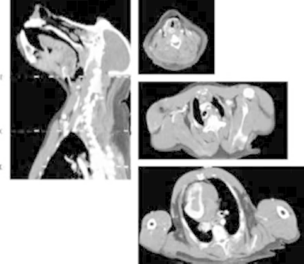
FIGURE 4.8
CT images of head, neck, and thorax (Papio anubis, adult female). (A) Mid-sagittal section of neck, deviating slightly laterally
through the cranial (superior) thorax; dotted lines refer to axial sections B, C, and D. (B) Axial section through the neck taken through the thyroid gland
and larynx at the level of the rima glottidis. Note the position of the carotid sheath and its neurovascular contents bilaterally adjacent to the laminae of the
thyroid cartilage. The laryngeal sac is a clearly visible radiolucency ventral to the thyroid laminae. (C) Axial section through the upper thorax. The well
developed pectoral muscles are seen crossing the anterior thorax to their insertions on the humerus. Note the dorso-ventral flattening of the trunk with
posterior orientation of the scapulae. Note also the axillary regions, well defined by their muscular borders, with the axillary vessels and cords of the
brachial plexus clearly seen on the right. (D) Axial section through the thorax at the hila of the lungs. The esophagus lies ventral to the vertebral column,
medial to the descending thoracic aorta. Note the narrow, elongated vertebral spinous process. (Images courtesy of Hansell Stedman.)
food in cheek pouches is not regurgitated material, but food
stored prior to mastication (
Lambert, 2005
).
fascia. This layer circumscribes the visceral compartment
by enclosing the pharynx and its caudal (inferior) exten-
sions: the esophagus and the respiratory assemblages of the
larynx and trachea (
Figure 4.8B
). Partially surrounding and
contiguous with these are the infrahyoid muscles, thyroid
and parathyroid glands, and the major neurovascular
structures of the ventral neck, including the innervation and
blood supply of local structures.
The muscles superficial to the visceral package are the
supra- and infrahyoid muscles. This series of muscles
Neck Viscera and Thyroid and Parathyroid
Glands
The visceral compartment of the neck in higher primates is
similar to that of humans in organization. Deep to the
investing fascia surrounding the sternocleidomastoid
muscle ventrally (anteriorly) and laterally is the pretracheal