Biomedical Engineering Reference
In-Depth Information
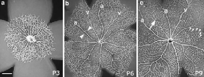
Fig. 4
Developing mouse retinal vasculature stained with an antibody against Collagen IV in
retinal whole mount preparations. The relatively uniform plexus visible in 3-day-old (P3) mouse
pups (
a
) expands and remodels by P6 (
b
) into a network with clearly distinguishable arteries (a)
and veins (v). Arteries can be identified by their capillary free zones (
arrowheads
in
b
). At P9
(
c
) the primary plexus has reached the retinal periphery, vessels start to sprout into the retina
and are visible as
white dots
(
small arrowheads
) establishing the deeper network of the retinal
vasculature. Some of the veins disappear from the primary plexus (
arrow
) and relocate by a process
of remodelling to the deeper plexus (not visible). Scale bar is 200
μ
m. Credits: [
18
]
A third step of the network formation is due to the vascular remodelling and
maturation.
Here we mainly consider the second step, during which the sprout of the network
starts due to the VEGF, and a planar network is formed, as in Fig.
4
.Themain
features of the model are the following. As already mentioned, the dynamics
involves three different
cell types
:
1. Type 1 cells: the
mural cells
which are the mature cells. They supply nutrients;
when a low concentration in their neighborhoods is detected (read, angioproteins
are present), they duplicate generating type 2 cells. Mural cells are subject to
death, while their displacement has been considered negligible.
2. Type 2 cells: the active cells, both the specialized
endothelial tip cells
at the
leading edge of the growing vascular network, and the
stalk cells
, located in the
neighborhood of tip cells. They can proliferate and die and their movement is
regulated by repulsive chemotaxis with respect to nutrients produced by type 1
cells and attractive chemotaxis with respect to the VEGF. When the type 2 cell
population increases, these cells start to organize, converting themselves into type
1 cells. Brownian diffusion may affect their movement.
3. Type 3 cells: the dead cells. Both type 1 and 2 may become type 3 cells.
The VEGF (
g
) and the nutrients (e.g., oxygen) (
u
) activate the migration and
the dynamics of endothelial cells at the microscale. We may suppose that at the
macroscale such fields may be described by continuum quantities evolving in time
via partial differential equations, whose parameters depend vice versa on the state
of the cells themselves.
Search WWH ::

Custom Search