Biomedical Engineering Reference
In-Depth Information
Radiometer) and a home-made working electrode made of pyrolytic graphite disks (4
mm diameter) and modified with the enzyme/ink layer.
The characterization of the optimized electrode was performed after replacing
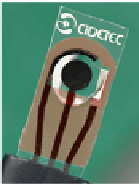
the previous system by carbon paste screen-printed electrodes (CPSPEs) with a three
electrode configuration (Fig. 1), including an Ag/AgCl pseudo-reference, a graphite
paste counter electrode and a graphite paste working electrode (3.1 mm diameter).
The CPSPEs were fabricated at CIDETEC facilities, as described by Ochoteco and
co-workers [17].
Fig. 1.
A screen-printed three-electrode system. (1) working electrode; (2) reference electrode;
(3) counter-electrode.
The one-compartment electrochemical cell containing 0.1 M KCl in 0.05 M Tris-HCl
buffer, pH 7.6 as supporting electrolyte, was thoroughly purged with Argon before each
experiment. Measurements were performed with a potentiostat Autolab PSTAT 12
(Eco-Chemie) monitored by the control and data acquisition software GPES 4.9. The
cyclic voltammograms (CV) were plotted at room temperature (22
C), with a scan
rate of 20 mV/s, in the potential window [0.0; -0.8] V (
vs
reference system). To evaluate
the biosensors response to the analyte (0.001 - 7 mM), the cell was successively spiked
with standard solutions of nitrite. After each addition, the electrochemical cell was
deoxygenated and the CV was registered. The catalytic currents (I
cat
) were measured at
the inversion potential (-0.8V); all values were subtracted from the non-catalytic current
recorded in the absence of nitrite (I
c
). Each experiment was replicated at least two times.
For amperometric measurements, the working potential was settled at -0.5 or -0.7 V,
with a speed rotation (
±
2
°
ω
) of 0 or 1000 rpm.
2.3
Bioelectrodes Preparation
Prior to coating, the pyrolytic graphite electrodes (PGEs) were polished with alumina
slurry in cloth pads. Then, the electrodes were thoroughly washed with DI water and
ethanol and ultrasonicated in water for 5 min. The electrodes surface was further
washed with DI water and dried with compressed air.
CPSPEs were used as produced, with no pre-activation. The conductive carbon
inks were previously diluted with an organic solvent (acetone or MEK) in a 1:1 ratio
and homogenised with the help of an ultrasound bath. The ink suspensions were then
mixed with ccNiR in different proportions (4:1, 2:1, 1:1 and 1:2 ink/enzyme). Finally,
a 5
L drop was placed on the surface of the working electrodes which were cured for
20 min. inside an oven set at 40°C or 60°C. Control experiments were carried out
with no curing treatment and/or no carbon ink; in such cases, the ccNiR layer was
dried at room temperature.
μ
Search WWH ::

Custom Search