Biomedical Engineering Reference
In-Depth Information
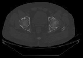
Fig. 1
The transverse section of total femur CT image
as acquired from the DICOM file images after conversion into MIMICS v10. Bone
tissue is then extracted by means of thresholding using default values that range
from 226 to 3,071 HU. The extracted bone tissue is put into a mask of volume
1,761,508.9874, 1,560,018.1976, and 1,841,722.1204 mm
3
, respectively, and the
number of pixels as 2,850,426, 1,984,073, and 3,089,900 pixels, respectively. These
pixels in the masks are modified using various tools successively: edit masks, region
growing, and calculation of 3D mask. Edit mask is used to separate the total femur
from the adjoining hard tissues like pelvis, tibia, etc. The region growing
tool provides the capacity to split the segmentation into separate masks with the
following properties:
Model 1—minimum -238 to maximum 1,695 HU, number of pixels 143,227, and
mask volume 88511.5585 mm
3
,
Model 2—minimum -150 to maximum 1,630 HU, number of pixels 492,348,
and mask volume 387118.7399 mm
3
,
Model 3—minimum -118 to maximum 1,744 HU, number of pixels 445,600, and
mask volume 265598.0377 mm
3
, respectively.
Calculate 3D from mask tool converts the 2-Dimensional images into 3D
models using an interpolation algorithm embedded in MIMICS as shown in Fig.
2
,
with the following properties of different models:
102228.88 mm
3
,
26172.09 mm
2
,
Model
1—mask
volume
surface
triangles
42,360, and points 21,230,
Model
471921.39 mm
3
,
134009.90 mm
2
,
2—mask
volume
surface
triangles
140,502, and points 69,989,
Model
328208.31 mm
3
,
115704.15 mm
2
,
3—mask
volume
surface
triangles
146,142, and points 72,871.
Search WWH ::

Custom Search