Biomedical Engineering Reference
In-Depth Information
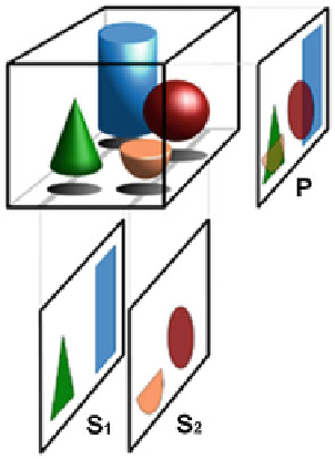
Fig. 1 Tomography
superposition with projected
image P
image produced is a tomogram. The method is used in radiology, archeology,
biology, geophysics, oceanography, materials science, astrophysics, quantum
information, and other sciences. In most cases, it is based on the mathematical
procedure called tomographic reconstruction. Figure
1
represents the basic princi-
ple of tomography; superposition-free tomographic cross sections S1 and S2
compared with the projected image P.
Medical Imaging-Perspective
The perspective in medical imaging is dominated by the development of newer
measuring technologies:
1. 3D tomography: In nondestructive material testing, 3D X-ray CT is widely used
in connection with a circular scanning geometry; i.e., the X-ray tube is moved
on a circle in a plane around the examined object with the detector plane
positioned at the opposite side. Helical geometry is favored in medical appli-
cations, but the necessary variable shift of the patient has not yet been solved
satisfactorily in existing algorithms. In principle, X-ray tube and detector can
be moved along arbitrary trajectories around the patient. The determination of
trajectories that are optimal with respect to resolution and stability remains a
mathematical challenge. Figure
2
a represents the X-ray source trajectory.
Figure
2
b and c is the mathematical representations of X-ray source trajectory.
2. Electron paramagnetic resonance imaging: In this technique, a decoupling of
the fourth dimension is not possible, since there, besides the three spatial
Search WWH ::

Custom Search