Biomedical Engineering Reference
In-Depth Information
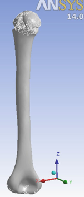
Fig. 5
3D Model for
analysis
area was bone tissue pixels, we defined this as a mask. Based on the mask
the humerus was selected, as in Fig.
2
. This mask is modified until we get
the satisfactory mask.
(b) Establishing FE model and generate surface mesh with MIMICS 10.01-
The generated region mask was used to develop 3D model for the bone.
Based on 3D Grayvalue interpolation techniques 2D images are trans-
formed into 3D model. It is a real 3D interpolation technique that takes into
account the Partial Volume effect and therefore it is more accurate [
16
]. To
obtain surface mesh, the option remeshing is used in order to raise the
quality of the triangles so that the preprocessor of an FEA package can
build a tetrahedron meshes from them (Fig.
3
).
(c) Convert surface mesh into volumetric mesh with ABAQUS 6.10-
This mesh is exported to ABAQUS 6.10 and is converted from tri into tetra
by using the mesh edit option. This is done because the denser the mesh the
more realistic and more accurate will be the solution.
Search WWH ::

Custom Search