Biomedical Engineering Reference
In-Depth Information
A
High/low
AB
AB
2
AB
4
B
AB
8
C
D
Low/high-high/low
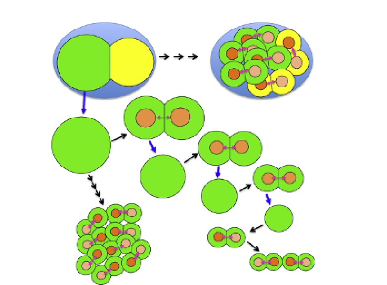
Figure 3.5 Regulation of POP-1 asymmetry in the AB lineage. In normal development,
(A) POP-1 asymmetry is first observed after the division of AB granddaughters (AB
4
).
(B) When P1 is isolated and cultured alone, POP-1 asymmetry is observed after the di-
vision of AB
4
, but the daughter proximal to the cell cluster tends to have higher POP-1
signal than the distal one that does not contact with the cluster. (C) The sequentially
isolated AB
8
cell shows POP-1 asymmetry. (D) In the next division, AB
8
daughters show
low/high
-
high/low POP-1 asymmetry. Green cells, pink double arrows, blue arrows, and
black arrows represent the AB lineage, daughter pairs, cell isolation, and cell divisions,
respectively.
embryo, tends to be enriched on the distal sides of cells, consistent with the
orientation of POP-1 asymmetry.
The POP-1 asymmetry was observed even in daughters of sequentially
isolated AB
4
cell but not in those of AB or AB
2
, indicating that cells can be
autonomously polarized without extrinsic Wnt signals after the AB
8
stage
(
Fig. 3.5
C). After the next division of sequentially isolated AB
4
cells,
POP-1 asymmetry in daughter pairs becomes opposite to each other with
low/high-high/low POP-1 localization which is not observed in normal
development (
Fig. 3.5
D). This observation together with higher POP-1
in proximal daughters in isolated AB (
Fig. 3.5
B) suggests that polarity ori-
entation is influenced by signals from or contacts with neighbor cells and that
this mechanism should be suppressed to orient polarity of all cells in the same
high/low POP-1 asymmetry during normal embryonic development.

