Biomedical Engineering Reference
In-Depth Information
A
E12
E13
E15
E10.75
E11.5
E17
C
B
KC
3
SCB
OHC
2
1
IHC
E
D
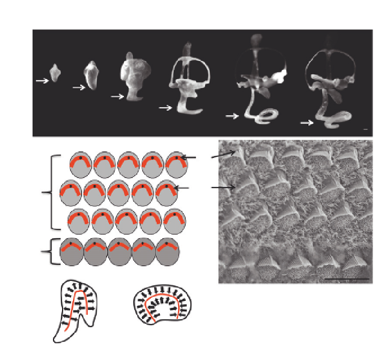
Figure 5.1 Development of the mammalian cochlea. (A) Lateral view of paint-filled
membranous labyrinths of embryonic mice at indicating ages between embryonic
day 10.75 and embryonic day 17. The cochlear duct (arrows) begins as a ventral out
pocketing that begins to extend from the otocyst at E10.75 and continues to extend
and coil until the early postnatal period. The mature mouse cochlear duct is between
6 and 10 mm and completes approximately 2.5 coils (scale bar is 100 mm; adapted from
Morsli, Choo, Ryan, Johnson, & Wu, 1998
). (B) Schematic representation of a surface view
of the organ of Corti. The single row of inner hair cells and three rows of outer hair cells
(OHC) are depicted, whereas the support cells that interdigitate between the hair cells
have been omitted. Each hair cell contains an actin-based stereociliary bundle, represen-
ted by a red arch, located on its lumenal surface. The bundles are uniformly oriented
toward the lateral edge of each cell. The microtubule-based kinocilium, represented
by a black dot, extends from the plasma membrane and is attached to the stereociliary
bundles via kinocilia links. (C) Scanning electron micrograph of the organ of Corti (basal
turn) at P0. The crescent-shaped stereociliary bundles (SCB) with the single kinocilium
(KC) located at the vertex are evident on both inner and outer hair cells. Microvilli on
the supporting cells between the hair cells are still present at this stage. Similarly, shorter
microvilli located on the medial surface of the hair cells are still present. (D and E) Sche-
matic representations of uniform stereociliary bundle orientations (arrows) in two ves-
tibular sensory epithelial, the saccule (D) and the utricle (E). Note the reversal zones (red
lines) present in each epithelium. Scale bar is 10 mm.