Biomedical Engineering Reference
In-Depth Information
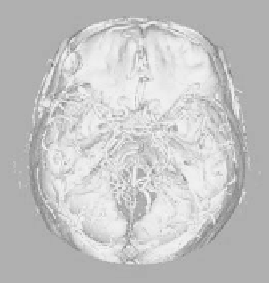
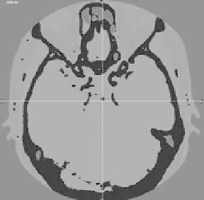
Figure 3.4 is a case where the blood vessel area is extracted by
the threshold processing by using commercial medical diagnosis
software. The shape of the brain blood vessel can be excellently
recognized from CT imaging as conirmed from this igure. However,
the separation of the blood vessels from the rest becomes dificult in
the area where the blood vessel and the bone approach each other.
This image conirms that blood vessels and bones have integrated
in the brainpan bottom, and that the CT image is suitable for the
areas where the blood vessel does not approach the bone. DSA,
which excellently extracts the blood vessel area, is achieved by
taking a luoroscopic image before and after the contrast medium
administration and operating the difference between them. It cannot
be inluenced by the existence of the bone area, and the shape of the
blood vessel can be extracted easier.
a)
b)
Figure 3.4
Reconstruction of three-dimensional vascular structure
based on individual image obtained by multi-slice CT
modality: (a) Extracted vessel area by threshold application.
(b) Reconstructed three-dimensional structure.
3.3.3
Three-Dimensional Vessel Shape Reconstruction
from MRI
In this section, we describe the re-composition of the three-
dimensional shape of the blood vessel using the cross-sectional data
of the head area obtained from MRI. In X-ray CT imaging, light and
shade are only decided depending on absorption (penetration) rate
of X-rays. On the other hand, for MRI, light and shade are generated
based on a signal depending on the density of atomic nucleus of


Search WWH ::

Custom Search