Biomedical Engineering Reference
In-Depth Information
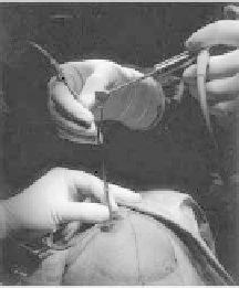
inner cylinder is inserted and implanted into the cerebral hematoma.
After that, the inner cylinder is removed and the aspiration tube (Fig.
2.19c) and the endoscope (Fig. 2.19d) are inserted. While inspecting
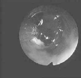
with the endoscope, the hematoma is suctioned (Fig. 2.20b) [51].
A case of left putaminal hemorrhage is shown in Fig. 2.21a. The
complete extraction of hematoma by endoscopic surgery is shown
in Fig. 2.21b.
Figure 2.20
Surgery: (a) Intra-operative environment appearance (b)
Endoscopic ield of view: In the back of the transparent sheath
a brown hematoma is pointed out with an arrow.
a)
b)
Figure 2.21
Head CT of left putaminal hemorrhage. (a) Before surgery
a relatively large hemorrhage is visible at the left putaminal
(arrow) (b) After surgery, almost all the hematoma was
extracted (arrow).


Search WWH ::

Custom Search