Biomedical Engineering Reference
In-Depth Information
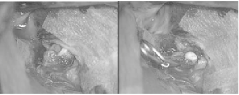
Figure 2.1
Clipping surgery for large unruptured Rt. MCA aneurysm: (a)
Pre-operative digital subtraction angiography image. A large-
size aneurysm is visible at Rt. MCA (*). (b) Intra-operative
photograph, the aneurysm is exposed (*). (c) Intra-operative
photograph, a clip was attached to the aneurysm neck. (d) DSA
image of the cerebral artery after the treatment.
a)
b)
c)
d)
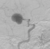
Figure 2.2
Clipping treatment for ruptured aneurysm in Rt. MCA (a,b) Pre-
operative 3D-CTA images. The arrow points to a hemorrhage
at the aneurysm in Rt. MCA. (c) Intra-operative photograph of
a subarachnoid hemorrhage. Red appearance is given by the
hemorrhage, the arrow points at a clot in the bleeding part of
the aneurysm. (d) Intra-operative photograph, the clip attached
to the aneurysm neck is pointed by the arrow.


Search WWH ::

Custom Search