Biomedical Engineering Reference
In-Depth Information
necessary to separate the CT value between the silicone elastomer
and contrast media, for instance, by adjusting the density of the
contrast medium, since these materials likely to take close CT value.
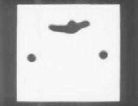
a)
b)
Figure 3.38
Fluoroscopic image of presented solid vascular model taken by
multi-slice CT imaging: (a) Cross-sectional image. (b) Three-
dimensional structure obtained by the reconstruction based
on a set of cross-sectional image.
3.11.5.2 Ultrasound imaging
Using the same experimental setup, the applicability of the presented
vascular model to ultrasound imaging was tested. Ultrasound imaging
was executed under this experiment composition by placing the
ultrasound probe tip near the vascular model, which is submerged
under water. To improve relection strength of the ultrasound wave
to the luid that is circulating inside vascular model, a small quantity
of aluminum powder was mixed in the circulating luid. Also to help
the transmission of the ultrasound wave inside the model, a special
jelly was spread over the model. Figure 3.39 shows the appearance
of the experiment. It was conirmed that the membranous vascular
model is applicable for this ultrasound imaging; on the other hand,
the solid vascular model is dificult applying this imaging modality
because the relection wave was very low as the transmission of
ultrasound wave in silicone elastomer was not good. Posterior tests
revealed that the single boundary offered by the solid coniguration
offers better imaging quality for intravascular ultrasound systems.
Chapter 7 shows how these models combined with intravascular
ultrasound systems are used for
in vitro
imaging of vasculature.
Figure 3.40 shows the ultrasound image obtained for the
membranous model of representative case No. 2. Figures 3.40a,


Search WWH ::

Custom Search