Biomedical Engineering Reference
In-Depth Information
of experimental setup for this low visualization experiment. To
remove the vascular deformation according to gravity and to
secure the visibility inside vessel lumen, the vascular model was
submerged under liquid, which has the same refraction index as the
vessel model. Moreover, the lumen of the vascular model was illed
with water solution that reproduces the viscosity of blood (glycerin:
33wt% and surface-active agent: 0.1wt%). Human blood stream was
reproduced with pulsatile blood pump (Harvard Apparatus, model
1405), which is commonly utilized in medical treatment. Here, to
reproduce the blood low rate of the basilar artery, a pulsatile blood
low of 200 mL/min with a beat rate of 75 was generated with the
pulsatile pump. And a pressure sensor (Fujikura Ltd., model-PSM)
was installed on the entrance part of arteria basilaris, to measure the
pressure transition during low visualization. The hypertension of
140
mmHg was set with this pressure sensor. Figure 3.41 shows the
low velocity wave on the entrance of arteria basilaris measured with
ultrasound sensor. This waveform indicates that human pressure
luctuation is satisfactorily reproduced.
Pulsatile Blood Pump
(Harvard corp. model-1405)
Dyestuff injection port
(hypodermic needle
No.27)
Inflow
Ultrasound Probe
(Not used for flow visualization)
Outflow
Inflow
Inflow
Cerebral artery
membranous model
Outflow
a)
Cerebral Arterial Model
b)
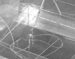
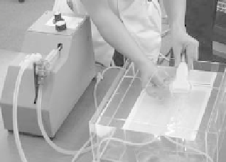
Figure 3.26
(a) Experimental setup for low visualization. (b) Dyestuff
injection into the vascular model for low visualization.
Under this experimental setup, a hypodermic needle No. 27
was introduced into the vessel lumen from the entrance part of
arteria basilaris, and its tip was ixed to the center of lumen. In this
state, above-mentioned water solution including white dyestuff
was injected into the low, and by this way, the blood stream was
made visible. Figure 3.26b shows the appearance at the dyestuff
injection. The details of the low in the BT aneurysm are shown in
Fig. 3.27, where the low is visualized for the solid vascular model
and membranous vascular model constructed for case No. 2.


Search WWH ::

Custom Search