Biomedical Engineering Reference
In-Depth Information
a)
b)
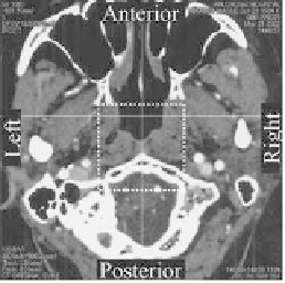
Figure 3.7
Head segment image obtained with CT angiography used
for reconstructing three-dimensional vascular lumen
coniguration; dotted line indicates the modeling area of 50
×
50
×
50 mm: (a) Transversal plane and (b) sagittal plane CT slice
images of the region of interest for vascular reconstruction.
a)
b)
Figure 3.8
(a) Three-dimensional igure of basilar artery with BT (basilar
tip) aneurysm, reconstructed from luoroscopic information.
(b) Vascular lumen model of basilar artery, fabricated by fused
deposition modality rapid prototyping (laminating pitch: 13
μ
m).
Then we arranged the above-fabricated vascular master mold
inside a cubic mold according to its orientation and embedded it
within liquid-state silicone rubber, which solidiies into transparent
elastomer through addition polymerization. Here, the physical
characteristics (elastic modulus, Poisson's ratio and friction
coeficient) of selected silicone are very similar to the arterial
tissue, and its excellent transparency allows easily observing the
endovascular intervention procedures. Here, molding the silicone into
cubic structure has the following advantages: It allows maintaining


Search WWH ::

Custom Search