Biomedical Engineering Reference
In-Depth Information
A
206
206
Nterm
Nterm
S1
SS3
S1
SS3
201
201
Y200
Y200
131
131
S
S
D
D
H
H
140
140
26
26
20
20
Cterm
Cterm
Trypsin = magenta
SMIPP-S-I1 = green
SMIPP-S-D1 = cyan
B
S1
S1
S
S
D
D
H
H
C
Y200
S
S
D
D
H
H
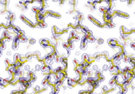
Figure 10.1
Overall structures of SMIPPs and comparisons with human trypsin. (A)
Cartoon representation showing structural alignment of SMIPP-S-I1
(green), SMIPP-S-D1 (cyan) and trypsin (1PTP; magenta), shown in stereo.
Regions of structural differences are indicated by residue labels. The loca-
tion of the disulphide bond (SS3) that is present in trypsin but absent in
SMIPPs is indicated. The side chain of Y200 which blocks the S1 subsite is
shown as stick bonds and labelled. (B) Structural differences in active site,
oxyanion (ellipse) hole and S1 subsite. (C) Electron density in active site,
contoured at 1s,wheres is the rmsd of electron density in the unit cell.