Biomedical Engineering Reference
In-Depth Information
Agonist Muscle
N
ag
F
p
F
ag
de
ac
F
gs
F
go
0
t
1
Time (s)
Antagonist Muscle
F
to
N
ant
ac
de
F
ts
F
ant
0
t
1
Time (s)
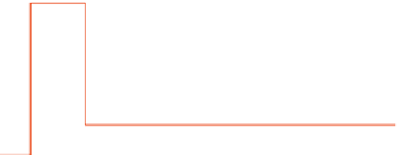
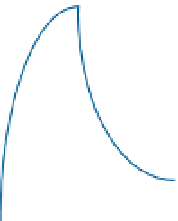
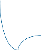
FIGURE 13.11
F
ant
,
active-state tensions. Note that the time constant for activation, t
ac
, is different from the time constant for deactiva-
tion, t
de
. The time interval,
Agonist,
N
ag
, and antagonist,
N
ant
, control signals and the agonist,
F
ag
, and antagonist,
t
1
, is the duration of the pulse.
follows most closely the neural input to the muscle. From Figure 13.11,apatternofneural
activity is observed as follows:
1.
The muscle that is being contracted (agonist) is stimulated by a pulse, followed by a step
to maintain the eyeball at its destination.
2.
The muscle that is being stretched (antagonist) is unstimulated during the saccade
(stimulated by a pause or a negative pulse to zero), followed by a step to maintain the
eyeball at its destination.
Figure 13.11 quantifies these relationships for the agonist neural
input,
N
ag
, and the
antagonist neural input,
N
ant
. The pulse input is required to get the eye to the target as soon