Biomedical Engineering Reference
In-Depth Information
fiber. In these sensors, light travels from a light source to the end of the optical fiber, where
it interacts with a specific chemical or biological recognition element. These transducers
may include indicators and ion-binding compounds (ionophores), as well as a wide variety
of selective polymeric materials. After the light interacts with the biological sample, it
returns either through the same optical fiber (in a single-fiber configuration) or a separate
optical fiber (in a dual-fiber configuration) to a detector, which correlates the degree of light
attenuation with the concentration of the analyte.
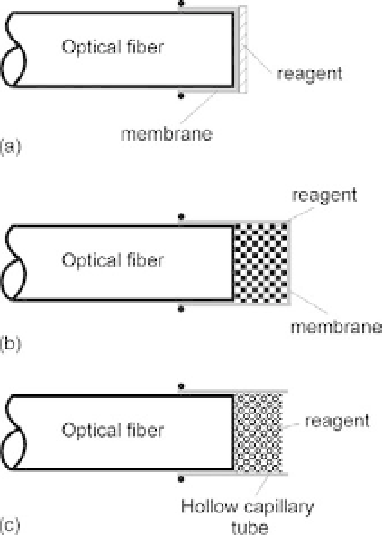
Typical indicator-mediated sensor configurations are shown schematically in Figure 10.41.
The transducing element is a thin layer of chemical material that is placed near the sensor
tip and is separated from the blood medium by a selective membrane. The chemical-sensing
material transforms the incident light into a return light signal with a magnitude that is pro-
portional to the concentration of the species to be measured. The stability of the sensor is
determined by the stability of the photosensitive material that is used and also by how effec-
tive the sensing material is protected from leaching out of the probe. In Figure 10.41a,the
indicator is immobilized directly on a membrane that is positioned at the end of the fiber.
An indicator in the form of a powder can also be physically retained in position at the end
of the fiber by a special permeable membrane, as shown in Figure 10.41b, or a hollow capil-
lary tube, as shown in Figure 10.41c.
10.6.7 Immunoassay Sensors
The development of immunosensors is based on the observation of ligand-binding reac-
tion products between a target analyte and a highly specific binding reagent. The key
FIGURE 10.41
Different indicator-mediated fiber optic sensor configurations.