Biomedical Engineering Reference
In-Depth Information
L Rectus Femoris
L Vastus Lateralis
L Vastus Medialis
L Hamstrings
L Anterior Tibialis
L Gastroc/Soleus
0
25
50
75
100
% Gait Cycle
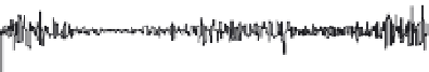
FIGURE 4.32
Electromyogram (EMG) data for the same cerebral palsy patient as in Figure 4.31. Plotted are EMG
activity signals for each of six left lower extremity muscles, each plotted as functions of percent of gait cycle. Gray bars
represent mean normal muscle activation timing.
flexed position throughout stance phase (0-60 percent of the gait cycle) when her foot is
contacting the floor. Knee motion in swing phase (60-100 percent) is also limited, with
the magnitude and timing of peak knee flexion in swing reduced and delayed. The range
of motion of her hip during gait is less than normal, failing to reach full extension at the
end of stance phase. The motion of her pelvis is significantly greater than normal, tilting
anteriorly in early stance coincident with extension of the hip, and tilting posteriorly in
swing coincident with flexion of the hip.
The deviations noted in these data illustrate neuromuscular problems commonly seen in
this patient population. Inappropriate hamstring tightness, observed during the clinical
examination, and inappropriate muscle activity during stance, seen in Figure 4.32, prevent
the knee from properly extending. This flexed knee position also impedes normal extension
of the hip in stance due to hip extensor weakness, also observed during the clinical exami-
nation. Hip extension is required in stance to allow the thigh to rotate under the advancing
pelvis and upper body. To compensate for her reduced ability to extend the hip, she rotates
her pelvis anteriorly in early stance to help move the thigh through its arc of motion. The
biphasic pattern of the pelvic curve indicates that this is a bilateral issue to some degree.
The limited knee flexion in swing combines with the plantar flexed ankle position to
result in foot clearance problems during swing phase. The inappropriate activity of the
rectus femoris muscle (Figure 4.32) in midswing suggests that spasticity of that muscle, a