Biomedical Engineering Reference
In-Depth Information
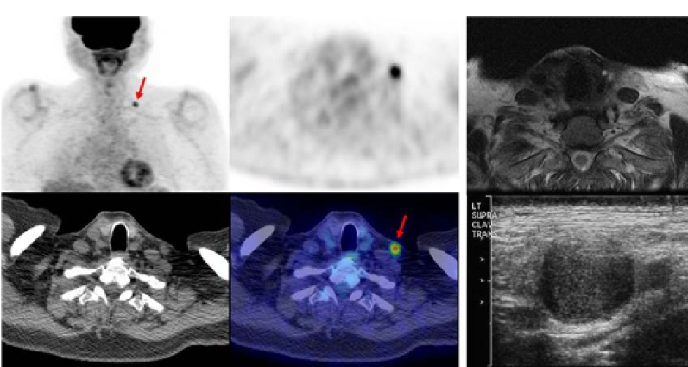
FIGURE 16.56
Images of different imaging modalities. (A) Upper left: PET frontal; (B) upper middle: PET
transverse; (C) upper right: MRI transverse; (D) bottom left: CT transverse; (E) bottom middle: PET/CT transverse
fused image; and (F) bottom right: ultrasound. Images of a 74-year-old male patient with nasal cavity esthesioneu-
roblastoma (a form of nasal cancer) who was restaged on a routine follow-up. PET identified a left supracalvicular
hypermetabolic nodal metastases (A, B, D, and E) that was not identified in the MRI (C). An ultrasound-guided
biopsy of the node was positive for esthesioneuroblastoma and then surgically resected (F).
Courtesy of Dr. Rathan
Subramaniam, 2010.
the PET and CT transverse, clearly emphasizes the metastases. For comparison, the MRI
image (C) of the same view, while providing spatial organization of the organs, is by itself
not as informative. Finally, the ultrasound image (F) provides a different view and was use-
ful for a guided biopsy.
16.8 SUMMARY
The different features and characteristics of three major imaging modalities are summa-
rized in Table 16.4 and shown in Figures 16.55 and 16.56. The tissue properties being
imaged are considerably different among the different modalities and can be complemen-
tary. Image fusion offers new opportunities to reconcile limitations of individual imaging
modalities. Different fusion combinations of imaging modalities have been attempted, but
PET/CT fusion is rapidly becoming the most popular [2]. Both CT and MRI can image
the whole body with consistent resolution and contrast; however, they are expensive meth-
ods of imaging and are slow and not portable. Ultrasound, on the other hand, has high but
variable resolution and penetration and limited access to certain portions of the body (intes-
tines, lungs, and bones). It is used to image soft tissues only, but it has the advantages of
low cost, portability, and real-time interactive imaging.