Biomedical Engineering Reference
In-Depth Information
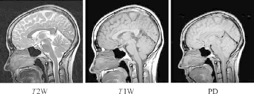
Different signal characteristics can be emphasized by a manipulation of the basic pulse
timing sequences (see Figure 16.47). For example, a short pulse repetition interval,
,
TR
and a short spin echo time,
, results in the commonly used
T
1
weighting (
1W) shown
TE
T
in Figure 16.50b. For example, typical values are
TE
¼
12 ms and
TR
¼
300 ms. Prolonging
TR
and the delays before and after
TE
produces a
T
2
-weighted (
T
2W) image as in
Figure 16.50a. An example of this is
TE
¼
100 ms and
TR
¼
3,000 ms. A combination of a
long
creates what is called a proton density weighted (PD) image, shown
in Figure 16.50c. For example, typical values are
TR
and a short
TE
TE
¼
30 ms and
TR
¼
3,000 ms. Recall that
T
1
is more sensitive to local thermal properties and
T
2
depends more on local transverse
magnetic field inhomogeneities. Many other alternative timing and pulse sequences are
possible and are useful for different clinical applications.
An important aspect of imaging is contrast among different types of tissues. In
Figure 16.51 longitudinal magnetization is shown for two tissues as a function of time,
indicating their different
T
1
rise times. Note that the contrast among the tissues varies
at different times, as indicated by the vertical separation between the curves at different
times. For example, compare the separations for times of 400 ms, 800 ms, and 1,600 ms.
Here it can be seen that contrast can be changed by timing intervals. Another way of
enhancing contrast
is to inject a paramagnetic medium or contrast agent such as
gadolinium.
16.3.8 Magnetic Resonance Imaging Applications
Functional MRI (fMRI) is the use of MRI to detect localized changes in brain activity,
usually in the form of changes in cerebral metabolism, blood flow, volume, or oxygenation
in response to task activation. These changes are interrelated and may have opposite effects.
For example, an increase in blood flow increases blood oxygenation, whereas an increase in
metabolism decreases it. The most common means of detection is measuring the changes in
the magnetic susceptibility of hemoglobin. Oxygenated blood is dimagnetic and deoxygen-
ated blood is paramagnetic. These differences lead to a detection method called blood
oxygen level dependent contrast (BOLD). Changes in blood oxygenation can be seen as
FIGURE 16.50
MR images of sagittal brain cross sections using three types of weighting planes: (a)
T
2
, (b)
T
1
,
and (c) proton density.
Courtesy of Philips Medical Systems.


