Biomedical Engineering Reference
In-Depth Information
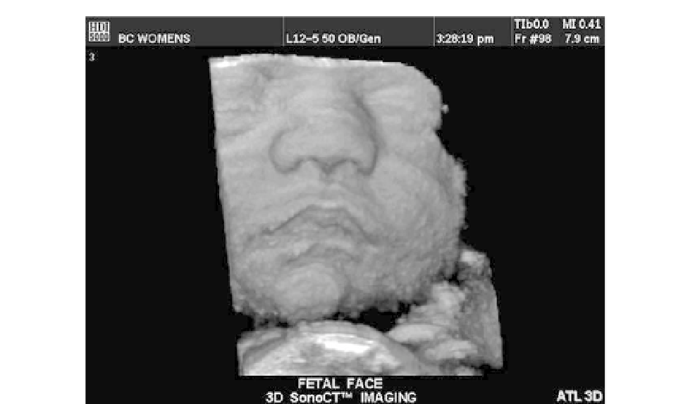
FIGURE 16.26
3D ultrasound image of a surface-rendered fetal head.
Courtesy of Philips Medical Systems.
When hydrogen is placed in a large static magnetic field, the magnetic moment of the atom
spins around it like a tiny gyroscope at the Larmor frequency, which is a unique property of
the material. For imaging, a radiofrequency rotating field in a plane perpendicular to the
static field is needed. The frequency of this field is identical to the Larmor frequency, and
once the atom is excited, the applied field is shut off and the original magnetic moment
decays to equilibrium and emits a signal. This voltage signal, detected by the same coils
used for the applied field, and two relaxation constants are sensed. The longitudinal mag-
netization constant,
T
1
, is more sensitive to the thermal properties of tissue. The transversal
magnetization relaxation constant,
T
2
, is affected by the local field inhomogeneities. These
constants are used to discriminate among different types of tissue and for image formation.
T
1
weighted images are used most often.
Today, MRI finds widespread application in the detection of disease and surgical
planning. MR images are highly detailed representations of internal anatomy. These may
be called parameterized images because considerable skill is involved in adjusting the
instrument to obtain images that emphasize different types of tissue contrast, the discrimi-
nation among different organ types and between healthy and pathological tissues. MRI is
used to examine most of the body, including the brain, abdomen, heart, large vessels,
breast, bones, as well as soft tissue, joints, cartilage, muscle, and the head and neck. It is
used for both children and adults and for detecting cancer pathologies, tumors, and
hemorrhaging.
An early precedent to MRI was nuclear magnetic resonance (NMR), first observed as a
phenomenon by Felix Bloch and Edward Purcell and their coworkers. They discovered that