Biomedical Engineering Reference
In-Depth Information
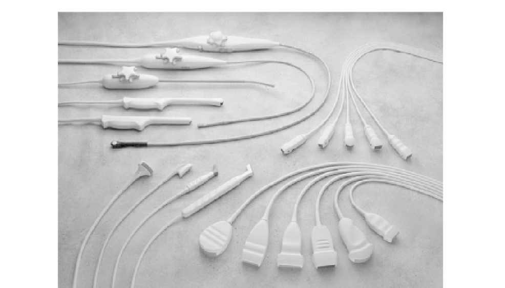
FIGURE 16.18
Transducers available on a modern imaging system are designed for a wide variety of clinical
applications. Transducer groups: bottom right: linear and curved linear arrays; top right: phased arrays; left side:
specialty probes including intraoperative, transesophageal, and transvaginal arrays.
Courtesy of Philips Medical
Systems.
to the body with a water-based gel. Except for regions containing bones, air, or gas, which
are opaque to imaging transducers, even small windows can be enough to visualize large
interior regions. The limitation of accessibility to viewing certain organs clearly is offset
by specialized probes such as transesophageal (down the throat) and intracardiac (inside
the heart) transducers that image from within the body.
16.2.4 Scattering
Interference of waves within the transducer plays an important role in the transduction
process. The crystal resonates when its thickness is half a wavelength, and the matching
layer is designed to be a quarter of a wavelength thick at resonance. The size of an object
relative to a wavelength is also a useful way of looking at acoustic scattering from objects.
Since ultrasound imaging is based on pulse-echoes returning from organs, it is necessary to
determine how the reflected signals are affected by the dimensions and shape of an object
relative to the insonifying wavelength.
Scattering falls within three ranges of effects: specular, diffractive, and diffusive
(Figure 16.19). Specular scattering is already familiar. When the dimensions of an object
are much greater than a wavelength, the reflected sound returns at the reflected angle
equal to the incident angle relative to the surface, as described in Section 16.2.2
on refraction. In this case, the reflected amplitude is determined by the reflection