Biomedical Engineering Reference
In-Depth Information
(c)
(d)
(a)
(b)
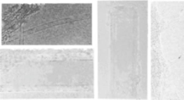
FIGURE 15.1
High-resolution transmission electron microscopy images of CNTs. (a) SWNT; (b)
MWNT; (c) closed MWNT tips (MWNT tips); and (d) closed SWNT tip. The separation between the closely
spaced fringes in the MWNT (b, c) is 0.34 nm, close to the spacing between graphite planes. The diameter
of the SWNT (a, d) is
1.2 nm. (Reprinted with permission from [8]. Copyright (1999) American Chemical
Society.)
FIGURE 15.2
High resolution STM image of the lattice structure of a helical semi-conducting SWNT.
(Reprinted with permission from [8]. Copyright (1999) American Chemical Society.)
collection of concentric SWNTs with different diameters. MWNTs may consist of one
up to tens and hundreds of concentric shells of carbons with adjacent shell separa-
tions of
0.34 nm. The carbon network of shells is closely related to the honeycomb
arrangement of the carbon atoms in the graphite sheets.
The diameters of CNTs range from 0.2 to 2 nm for SWNTs and from 2 to 100 nm
for MWNTs, while the lengths of CNTs range from several hundred nanometers to
several micrometers [13, 16]. The structural information of CNTs has been provided
by both transmission electron microscope (TEM) and scanning tunneling microscopy/
spectroscopy (STM/STS) at the atomic level. Figure 15.1 shows TEM images of both
SWNTs and MWNTs. In these images obtained by high resolution TEM, the diameter
of the SWNTs of
1.2 nm is clearly seen. The spacing (0.34 nm) between graphite
planes of MWNTs as well as the closed nanotube tips is also seen in the TEM images.
Atomically resolved lattices of SWNTs have been observed by STM as shown in
Fig. 15.2. This STM image also shows the helicity of SWNTs. For the formation of
curved structures such as the tips of CNTs and fullerenes from a planar hexagonal
graphite lattice, certain topological defects and pentagons (12 pentagons are needed to
close the hexagonal lattice) need to be introduced.


Search WWH ::

Custom Search