Biomedical Engineering Reference
In-Depth Information
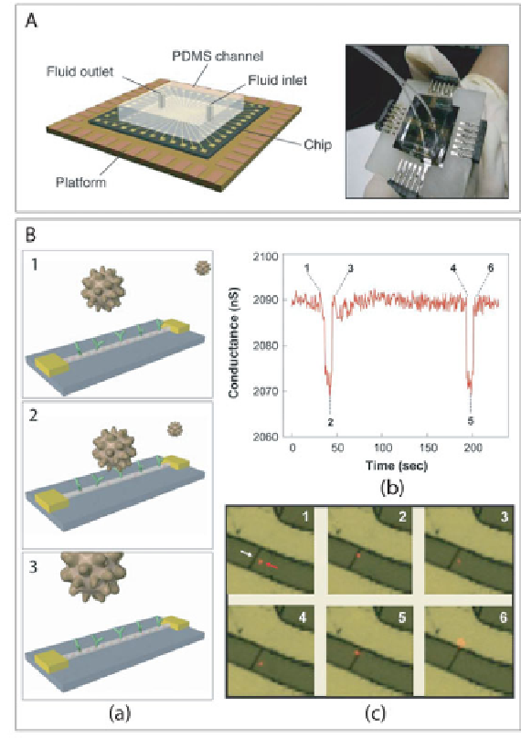
Figure 2. A)
(left) Schematic and (right) photograph of a prototype nanowire
sensor biochip with integrated microfluidic sample
delivery. B) (a) Schematic
illustration of a single virus binding and unbinding to the surface of a NWFET
modified with antibody receptors. (b) Conductance vs. time data recorded from a
single device modified with anti-influenza type A antibody. (c) Optical data rec-
orded simultaneously with conductance data in (b). Combined bright-field and
fluorescence images correspond to time points 1-6 indicated in the conductance
data; virus appears as a red dot in the images.
Search WWH ::

Custom Search