Biomedical Engineering Reference
In-Depth Information
(a)
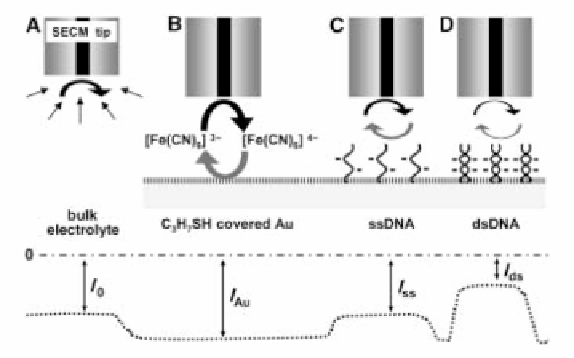
Figure 9. (a) Schematic representation of repelling-mode SECM. A) A diffu-
sion-limited current flows due to the reduction of [Fe(CN)
6
]
3-
at a tip potential of 0
mV (vs. Ag/AgCl in 3M KCl). B) Recycling of tip-generated [Fe(CN)
6
]
4-
at the
propane thiol-modified Au surface causes positive feedback. C) Above a DNA spot,
tip-generated [Fe(CN)
6
]
4-
hindered to diffuse into of the Au surface due to the elec-
trostatic repulsion. D) Upon hybridization, mass transport is further suppressed due
to the increasing degree of electrostatic repulsion that further increases. (b)
Three-dimensional SECM image of an individual spot of a single stranded 20-base
oligonucleotide as obtained by the repelling-mode SECM in 5 mM [Fe(CN)
6
]
3-
in
0.1 M phosphate buffer (pH 5.7) containing 3 M NaCl. For the tip electrode a
10-μm-diameter Pt UME was used and its electrode potential was fixed at 0 mV for
[Fe(CN)
6
]
3-
reduction. (c) Detection of hybridization through electrostatic repulsion
and visualization of DNA duplex formation by means of repelling-mode SECM. A
selected spot of a 20-base oligonucleotide was exposed to the hybridization buffer
(1 M NaCl-0.1 M phosphate buffer, pH 6.3) containing 2 μM of the complemen-
tary oligonucleotide target. A neighboring spot was subjected to target-free hybrid-
ization buffer (control). SECM line scans were measured by operating the tip of a
10-μm-diameter Pt microelectrode (0 mV vs. Ag/AgCl in 3M KCl) in solutions of 5
mM [Fe(CN)
6
]
3-
in 3 M NaCl-0.1M phosphate buffer (pH 5.7). All pictures were
reprinted from Ref. 39, Copyright (2004) Wiley-VCH Verlag GmbH& Co. KGaA.
Reproduced with permission..
Search WWH ::

Custom Search