Biomedical Engineering Reference
In-Depth Information
12 nA 16 nA
300
P
m
0 700
P
m
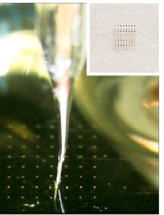
Figure 7. An example of SECM imaging of microe-
lectrode array. Top: Setup of the electrochemical
cell: An UME (Pt,
r
= 10 μm) approaches an 8 u 8
array electrode consisting of each 50-μm diameter
Au-UME that was embedded in a glass substrate
with 500-μm spacing. Inset shows a top-view of the
array electrode. Bottom: A representative current
mapping image on an arbitrarily chosen two elec-
trodes. The entire electrode setup was soaked in an
electrolyte solution containing 10 mM K
4
[Fe(CN)
6
]
(0.1 M KCl) as mediator. The electrode potential of
the tip was kept at +0.6 V (Ag/AgCl) for mediator
oxidization while those for the target electrodes were
left at their open-circuit potentials. Changes of the
oxidation current evolved with raster scan of the tip
were measured and encoded into each pixel.


Search WWH ::

Custom Search