Biomedical Engineering Reference
In-Depth Information
4 μm
20 μm
Blood vein
(a)
(b)
6 μm
6 μm
Blood vein
Blood vein
(c)
(d)
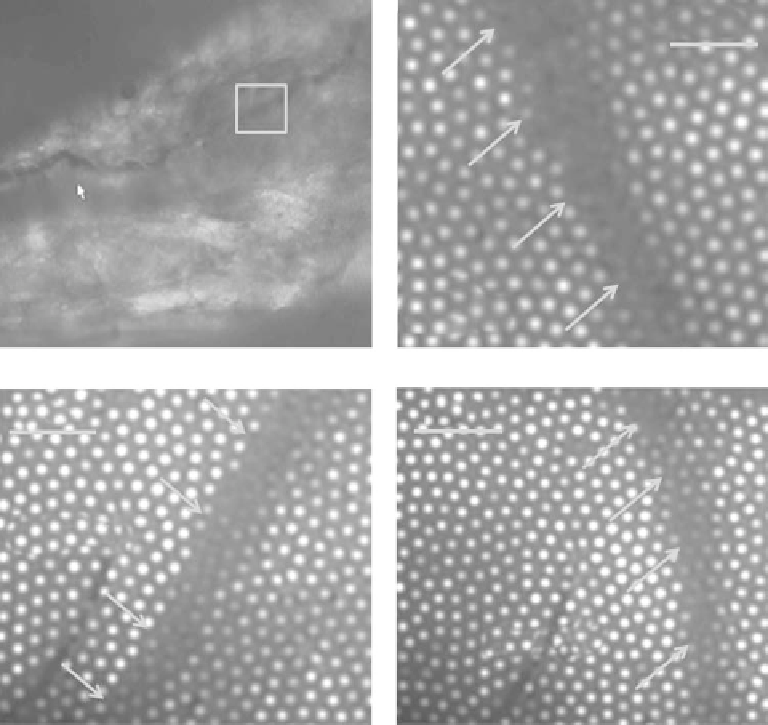
Figure 26.8
(See companion CD for color igure.)
.Blood.veins.inside.a.chicken.tissue..(a).Top.view.microscope.image..(b-d).
Imaging.of.the.chicken.wing.veins.using.the.special.micro.probe.
isolated.as.described.in.Fixler.et.al..(2002)..Briely,.hearts.from.newborn.rats.were.rinsed.
in.phosphate.buffered.saline.(PBS),.cut.into.small.pieces.and.incubated.with.a.solution.of.
proteolytic. enzymes-RDB. (Biological. Institute,. Ness-Ziona,. Israel).. Separated. cells. were.
suspended.in.Dulbecco's.Modiied.Eagle's.Medium.(DMEM).containing.10%.inactivated.
horse. serum. (Biological. Industries,. Kibbutz. Beit. Haemek,. Israel). and. 2%. chick. embryo.
extract,.and.centrifuged.at.300.×.g.for.5.min..Precipitant.(cell.pellet).was.resuspended.in.the.
growth.medium.and.placed.in.culture.dishes.on.collagen/gelatin.coated.cover-glasses..On.
day.4,.cells.were.treated.with.0.5-5.μM.of.DOX.for.18.h.and.then.with.drug-free.growth.
medium.for.an.additional.24.h..Cell.samples.were.grown.on.microscope.cover.slips.and.
imaged.by.the.micro.probe.
Figures.26.8.and.26.9.present.imaging.of.blood.veins.inside.a.chicken.wing..Figure.26.8a.
shows. top. view. microscope. image. of. the. veins. of. the. chicken. wing. area,. while. Figure.
26.8b.through.d.show.these.veins.(indicated.by.the.solid.arrows).imaged.with.the.special.
micro.probe.