Biomedical Engineering Reference
In-Depth Information
158
5 μm
200 nm
10
(b)
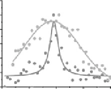
Cofocal (237 nm)
STED (39 nm)
1.2
1.0
0.8
0.6
0.4
0.2
0.0
y
2 μm
x
0
100
200
300
400
44
3303
Position/nm
(a)
(c)
Figure 25.9
(See companion CD for color insert.)
.Confocal.and.STED.imaging.of.HeLa.cells.labeled.with.BSA-conjugated.
FNDs. by. endocytosis.. (a). Confocal. image. acquired. by. raster. scanning. of. an. FND-labeled. cell.. The. luo-
rescence. image. of. the. entire. cell. is. shown. in. the. white. box. demonstrating. fairly. uniform. cell. labeling. by.
BSA-conjugated. FNDs.. (b). STED. image. of. single. BSA. conjugated. FND. particles. enclosed. within. the. green.
box. in. (a).. (c). Confocal. and. STED. luorescence. intensity. proiles. of. the. particle. indicated. in. (b). with. a. blue.
line..Solid.curves.are.best.its.to.1D.Gaussian.(confocal).or.Lorentzian.(STED).functions..The.corresponding.
full.widths.at.half-maximum.are.given.in.parentheses..(From.Tzeng,.Y.-K.,.Faklaris,.O.,.Chang,.B.-M.,.Kuo,.Y.,.
Hsu,.J.-H.,.and.Chang,.H.-C.:.Superresolution.imaging.of.albumin-conjugated.luorescent.nanodiamonds.in.
cells.by.stimulated.emission.depletion..
Angew. Chem. Int. Ed
..2011..50..2262-2265..Copyright.Wiley-VCH.Verlag.
GmbH.&.Co..KGaA,.Reprinted.with.permission.)
Most. recently,. Tzeng. et. al.. have. applied. the. STED. technique. to. study. the. homogenous.
labeling. of. HeLa. cells. with. 30.nm. FNDs. (Tzeng. et. al.. 2011).. The. FND. particles. are. irst.
coated. with. BSA. non-covalently. to. prevent. agglomeration. in. cell. medium. and. then.
delivered.to.the.cell.cytoplasm.by.endocytosis..With.STED,.the.authors.have.been.able.to.
identify.individual.FND.particles.in.cells.and.distinguish.them.from.particle.aggregates.
trapped.in.endosomes.(Figure.25.9).
Another. super-resolution. imaging. technique. applicable. to. FND. is. known. as. ground.
state. depletion. (GSD). microscopy.. The. technique. takes. advantage. of. the. intermediate.
state,.
1
A,.of.the.(N-V)
−
.center.(Rittweger.et.al..2009b)..The.idea.is.that.when.the.excitation.
laser. power. is. high. enough. to. saturate. the. transition.
3
A.→.
3
E,. the. accumulation. of. the.
populations.in.the.intermediate.state.will.switch.off.the.luorescence.from.
3
E→.
3
A..Similar.
to.STED,.the.GSD.microscopy.utilizes.a.doughnut-shaped.high-power.laser.beam.and.a.
co-aligned.excitation.laser.beam..The.doughnut-shaped.beam.excites.the.(N-V)
−
.center.to.
the.intermediate.state,.resulting.in.depletion.of.the.ground.state.populations.and.therefore.
the.
3
E.→.
3
A.luorescence..Rittweger.et.al..have.applied.the.technique.to.improve.the.spatial.