Biomedical Engineering Reference
In-Depth Information
y
=
h
F
DEP
F
Stokes
F
Gravity
v
=max@y =
h
/2
Hydrodynamic velocity
profile (
v
)
F
DEP
F
Stokes
y
x
F
Gravity
y
= 0
(a)
Electrode arrays
-veDEP
Flow
+veDEP
-veDEP
(b)
100
80
60
40
20
0
0
0.5
1.5
Distance along electrode array (mm)
1
2
2.5
3
3.5
(c)
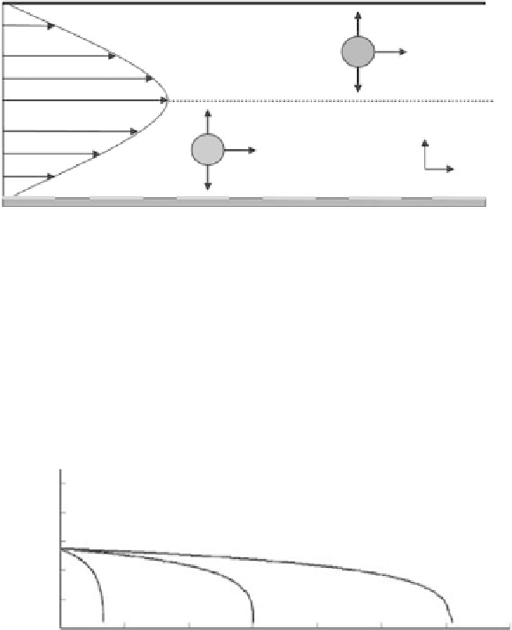
Figure 17.12
(See companion CD for color igure.)
. DEP-based. ield. low. fraction.. (a). Schematic. diagram. of. the. DEP-FFF.
geometry. and. the. involved. physical. forces,. (b). Particle. focusing. and. fractionation. mechanism,. and. (c). the.
simulated.trajectories.of.monocytes,.B.lymphocytes,.and.T.lymphocytes..(Reproduced.with.permission.from.
Holmes,.D.,.Green,.N.G.,.and.Morgan,.H.,.Microdevices.for.dielectrophoretic.low-.through.cell.separation,.
IEEE
Eng. Med. Biol.,
.22,.85-90,.Copyright.2003,.IEEE.)
As.shown.in.Figure.17.12a,.the.interactions.of.DEP.force,.gravity,.and.the.viscous.drag.force.
decide.the.velocity.of.particles.and.their.steady-state.locations.above.the.electrode.surface.
(Hughes.2002b)..When.the.forces.are.in.equilibrium,.the.following.relationship.is.held:
2(
ρ
−
ρ
)
g
( )
∇ =
p
m
2
Re
[
K
ω
]
E
.
(17.14)
3
ε
.
m
From.Equation.17.14,.the.location.of.a.particle.can.be.levitated.in.DEP-FFF,.depending.on.
their.density,.permittivity,.and.the.received.electric.signal..Due.to.the.parabolic.velocity.
proile. of. the. hydrodynamic. low,. such. levitation. varies. across. the. channel. height,. and.