Biomedical Engineering Reference
In-Depth Information
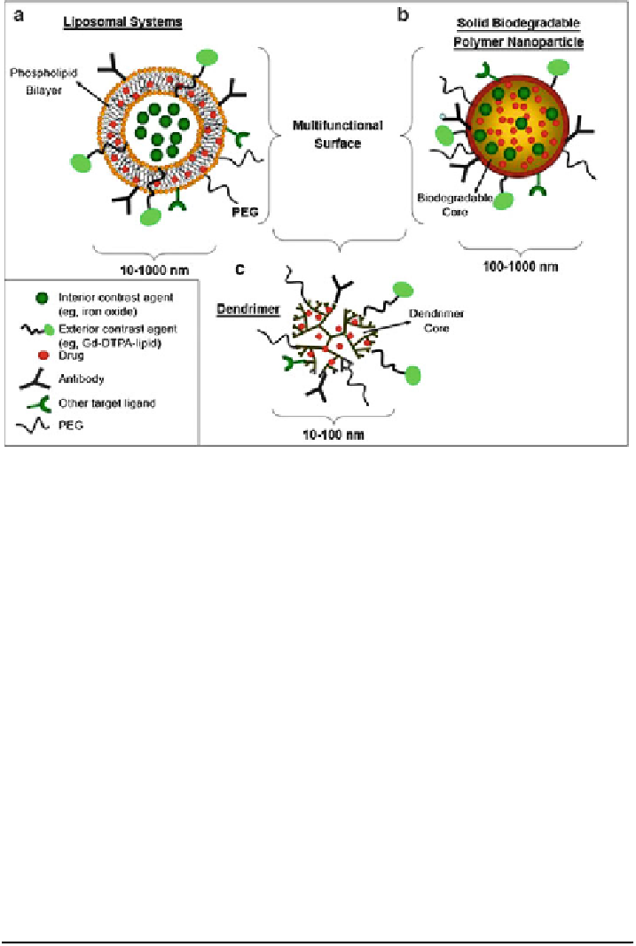
Fig. 8 Nanosystems that may function as simultaneous drug delivery and imaging agents for
targeting T cells: (a) liposomal systems, (b) solid biodegradable nanoparticulates, and (c) macro-
molecular dendrimer complexes.
PEG
polyethylene glycol,
Gd-DTPA
gadolininum-diethylene
triamine penta acetic acid. (Adapted from [
48
])
Syto16 dye solution for 10 min followed by a 1 nM of QD605-AST1 solution for
30 min. In Fig.
7
a we see that the cell nucleus is preferentially stained green by
Syto16 due to its cell permeability and DNA intercalation. The yellow-orange color
indicates colocalized CdSe
ZnS QD-AST1 and Syto16). Phase, fluorescence, and
overlay images of A431 and 3T3 cells were labeled using solutions of 0.5 nM QD-
streptavidin) and 0.5 nM CdSe
ZnS QD-AST1 (Fig.
7b-e
) Some different
strategies for nanosystems are illustrated in Fig.
8
. Figure
8
a shows liposomal
systems, Fig.
8
b solid biodegradable nanoparticles, and Fig.
8
c macromolecular
dendrimer complexes.
6.1.3 Aptamer-Conjugated Nanoparticles
Recently, nanoparticle-aptamer bioconjugates, as shown in Fig.
9
, have been
developed and demonstrated for targeted delivery to cancer cells. The imaging
Fig. 7 (continued) color indicates colocalized QD-AST1 and Syto16. (b-e) Phase, fluorescence,
and overlay images of A431 (b, c) and 3T3 (d, e) cells labeled using solutions of 0.5 nM
QD-streptavidin (b, d) and 0.5 nM QD-AST1 (c, e).
Scale bars
:25
m
m. (Adapted from [
47
])