Biomedical Engineering Reference
In-Depth Information
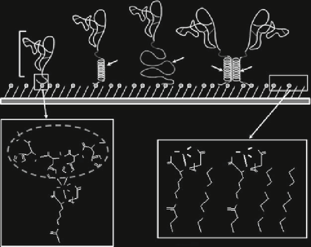
EGF-His
EGF-K5-His
EGF-E5-His
dEGF-His
EGF
E5
K5
K5
E5
Gold-evaporated glass plate
Oligohistidine
sequence
H
N
O
H
O
N
O
O
O
O
O
H
O
O
Ni
Ni
N
O
O
N
N
H
N
O
H
N
N
N
N
O
O
OH
OH
OH
N
N
N
O
N
O
N
O
O
O
Ni
O
O
O
O
O
N-H
N-H
N
O
O
O
O
O
O
N-H
O
Ni-NTA andTEG-thiol mixed SAM
His - Ni-NTA
Fig. 6 EGF-containing chimeric proteins anchored to the Ni-chelated surface through coordina-
tion.
Bold lines
in the molecular structures represent chelate bonding.
TEG-thiol
triethylene
glycol-containing alkanethiol. Reproduced from Nakaji-Hirabayashi et al. [
87
] with permission
from American Chemical Society, copyright 2009
3.3 Proliferation of Rat NSCs on EGF-His-Immobilized Surface
We investigated the efficiency of NSC expansion on surfaces with EGF-His
immobilized in the correct orientation. NSCs were obtained from neurosphere
cultures prepared from fetal rat striatum harvested on embryonic day 16. NSCs
were cultured for 5 days on EGF-His-immobilized substrates prepared with mixed
SAMs of different COOH-thiol contents. Cells adhered and formed network
structures at a density that increased with the COOH-thiol content of the surface.
As a control, cells were seeded onto surfaces without immobilized EGF-His. This
resulted in poor cell adhesion during the entire culture period. In addition, when
EGF-His adsorbed to SAMs with 100% COOH-thiol or SAMs with NTA-
derivatized COOH that lacked Ni
2+
chelation, we observed poor initial cell adhe-
sion, and the cells formed aggregates within 5 days. Interestingly, the substrate used
to covalently immobilize EGF-His with the standard carbodiimide chemistry was
not a suitable surface for cell adhesion and proliferation. The control experimental
results contrasted markedly with results from EGF-His-chelated surfaces.