Biomedical Engineering Reference
In-Depth Information
a
CCD camera
Emission filter
Objective lens
Glass window
Teflon borrel
Slilcone rubber
spacer
Substrate
q
Laser
(
λ
= 532 nm)
Prism
b
(1)
(2)
(3)
(4)
Cell
Evanescent
field
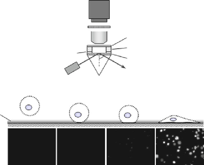
Fig. 1 Real-time tracking of cell adhesion [
42
]. (a) Components of a total internal reflection
fluorescent microscope (TIRFM). (b) The cell adhesion process; (
1
) a cell approaches the surface,
(
2
) the cell lands, (
3
) the cell attaches, and (
4
) the cell spreads out on the surface. The evanescent
field was generated by total internal reflection of a laser beam at the glass-water interface. Cells
with fluorescently labeled membranes (
dashed lines
) were plated on SAMs. Cell membranes
within the evanescent field (
solid line
) were observed by TIRFM. Corresponding TIRFM images
are shown
below
real-time tracking of cell behavior. To observe initial cell adhesion onto SAMs in
real
time, we employed a total
internal
reflection fluorescence microscope
(TIRFM).
The optical assembly of a TIRFM is schematically shown in Fig.
1a
[
42
].
The TIRFM utilizes an evanescent field, which is generated by laser reflection at
a water-glass interface [
43
,
44
]. The intensity of the evanescent field decays
exponentially with increasing distance from the interface. The characteristic
penetration depth of the evanescent field is approximately 100 nm (depending on
wavelength, incident angle, and refractive index of the glass and aqueous solution).
Fluorescent dyes are excited in the evanescent field, and the emitted fluorescent
light is captured with a charge-couple device (CCD) camera.
We assembled a TIRFM with low magnification to study cell adhesion behavior
on SAMs with various functional groups [
42
]. Figure
1b
shows a schematic
illustration of the cell adhesion process and the corresponding TIRFM images.
A suspension of cells with fluorescently labeled cell membranes is applied
onto a substrate (Fig.
1b
-1). At first, no bright spots were observed by TIRFM,