Biomedical Engineering Reference
In-Depth Information
and potential biologic beneits associated with the CaP crystals may
play a key role in enhanced site response, potentially improving
clinical predictability and outcomes. Preclinical studies demonstrate
a substantial improvement on the rate and extent of osseointegration
for the NanoTite Implant versus the Osseotite Implant.
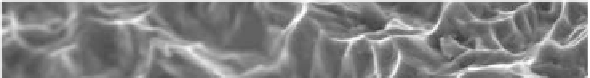
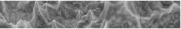
Figure 13.7
OSSEOTITE
®
Surface at 20,000× [http://biomet3i.com].
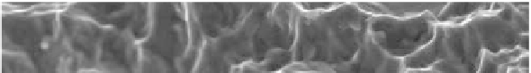
Figure 13.8
NanoTite™ Surface at 20,000× [http://biomet3i.com].
The nanoscale CaP surface created by DCD (Nanotite, 3i) was
evaluated [39]. The histologic evaluation of clinical implants
revealed bone-to-implant contact of 19 ± 14.2% and 32.2 ± 18.5%
for the Osseotite (3i) control and the Nanotite (3i) experimental
implants, respectively. Additionally, greater bone formation at 4
and 8 weeks was observed [19].
Recently, ion-beam assisted deposition (IBAD) process, which
provides increased integration with the implant surface, known
as high-energy sputter deposition, has been used to create a
commercially available dental implant surface [6]. In the NanoTite™