Biomedical Engineering Reference
In-Depth Information
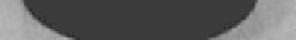
Figure 10.10
Carbon nanotube powders (a) and the sintered carbon
nanotube monolith (b) [61].
Table 10.2
Comparison of the properties of CNTs monolith with bone
[6, 61]
Yo u n g
modulus
(GPa)
Compressive
strength
(MPa)
Flexural
strength
(MPa)
Bulk density
(g/cm
3
)
Vickers
hardness
Material
Bulk CNTs 1.95
44
20
249
172
Bone
1.9
<60
19
150
180
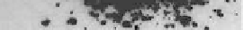
The
in vivo
reactions to the CNT monolith reveals that 1 week
after implantation, the CNT monolith was surrounded by tissue
with many cells like ibroblasts, ibroblasts with spindle-shaped
cytoplasm, and some inlammatory round cells (Fig. 10.11a). 4
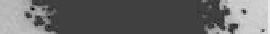
weeks after implantation, the CNT monolith was covered by loose
ibrous connective tissue, and inlammation around materials
was slight in comparison to that after 1 week (Fig. 10.11b). No
severe inlammation such as necrosis, degeneration, or neutrophil
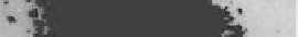
iniltration was observed around the CNT materials [61]. At 1
week after implantation in the femur, active callus formation from
the periosteum and immature newly formed bone were observed
around the CNT monolith. The newly formed bone did not directly
attach to the material (Fig. 10.11c). At 4 weeks, newly formed bone