Biomedical Engineering Reference
In-Depth Information
The knowledge of the anatomical shape of the crowns of the
teeth is the basis of the choice selection of prosthetic reconstructions
of the missing teeth in patients. The position of the teeth in the arch
is in respect to their function, meaning that they are supported
in the alveolar sockets through a connective tissue called the
periodontal ligament to play a distinctive role in the mastication
process. The main function of the periodontium is to attach the
teeth to the bone and to maintain the integrity of the surface of the
masticatory mucosa of the oral cavity [5]. Also called “attachment
apparatus,” it undergoes many changes with age and also due to
functional and oral environment alterations.
Maxillary teeth axes lie oblique to the vertical axis of the cranium.
Roots in the dental arch in the maxilla are more closely spaced than
are the crowns of the teeth, which appear to slightly tilt outward. As
goes for the axes of the mandibular teeth, they are inclined inward
relative to the vertical axis of the cranium, such that their crowns on
the opposite side of the jaw lie closer than the roots.
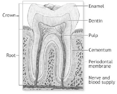
The anatomical crown of the tooth is covered by enamel, a highly
mineralized tissue, up to the cervical region of the tooth (Fig. 2.6).
Underneath lies the dentin, which forms the main core of the tooth.
This structure contains 70% of inorganic substance, making it
less brittle from the enamel. In the middle of the tooth is the pulp
chamber containing vessels and nerves.
Figure 2.6
The anatomy of the tooth.