Biomedical Engineering Reference
In-Depth Information
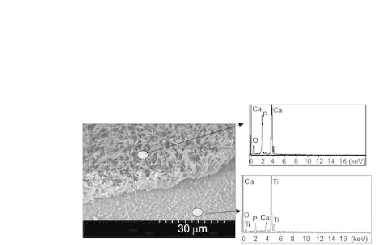
The EDS analysis of the surface after Ca-P removing shows Ca
and P components (Fig. 9.83), and Raja
et al
. suggest that calcium
phosphate grows from the bottom of the nanotubes rather than
just sticking on the top surface of the titanium oxide [75]. Calcium
phosphate coating on a polished lat titanium surface is extremely
weak, not uniform and is easily removed after washing [75].
Figure 9.83
EDS analyses of the tensile tested surface where the coating
was partially removed. The upper spectrum is for the Ca-
P coating. The lower spectrum is for the surface, where the
coating was removed (the peaks of Ca and P are still observed,
indicating that the calcium phosphate nucleated at the bottom
of the nanotubes) [75].
References
1. Anselme, K., Linez, P., Bigerelle, M., Le Maguer, D., Le Maguer, A.,
Hardouin, P., Hildebrand, H.F., Iost, A., and Leroy, J.M. (2000). The relative
inluence of the topography and chemistry of Ti-Al6-V4 surfaces
on osteoblastic cell behaviour,
Biomaterials
,
21
, pp. 1567-1577.
2. Balasundaram, G., Sato, M., and Webster, T.J. (2006). Using hydroxy-
apatite nanoparticles and decreased crystallinity to promote osteoblast
adhesion similar to functionalizing with RGD,
Biomaterials
,
27
,
pp. 2798-2805.
3. Ban, S., Iwaya, Y., Kono, H., and Sato, H. (2006). Surface modiication
of titanium by etching in concentrated sulfuric acid,
Dental Materials
,
22
, pp. 1115-1120.
4. Bauer, S., Kleber, S., and Schmuki, P. (2006). TiO
2
nanotube: tailoring
the geometry in H
3
PO
4
/HF electrolytes,
Electrochem
.
Commun
.,
8
,
pp. 1321-1325.