Biomedical Engineering Reference
In-Depth Information
sides of the skull. The masseter, temporalis, and medial pterygoid
serve to elevate the mandible when they constrict (Fig. 2.3). As goes
for the lateral pterygoid, when shortening in conjunction with other
muscles, as well as the opposite lateral pterygoid, serves to depress,
protrude, or shift the mandible laterally.
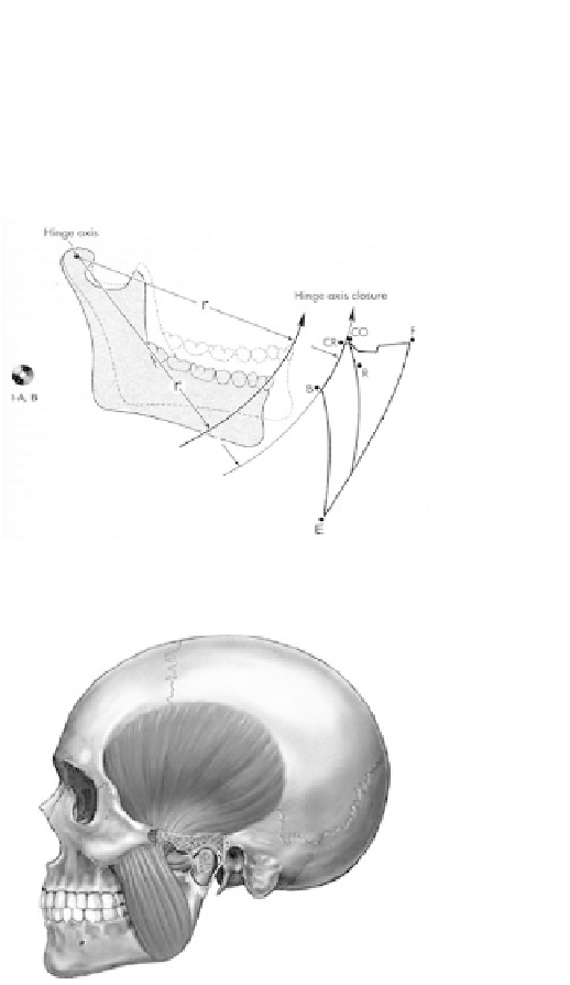
Figure 2.2
Mandibular movement projected on saggital plane.
Figure 2.3
Human skull showing masseter and temporalis muscles.
Moreover, the suprahyoid muscles extending from the skull and
mandible to the hyoid bone serve to depress the mandible, strain
loor of the mouth and cooperate in deglutination. The muscles of
facial expression are too weak to be involved in the movement of
mandible; however, they play a vital role in the esthetics.