Biomedical Engineering Reference
In-Depth Information
content existed on the anodized Ti specimen with respect to the
untreated Ti specimen [112]. The results are consistent with those
reported by Woo
et al
. [104]. They found that scaffolds with nano-
ibrous pore walls adsorb more proteins than scaffolds with solid
pore walls [104]. The nanoscale topography surface enhances the
cell adhesion of ibroblast cells.
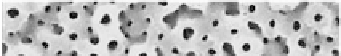
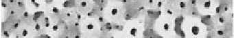
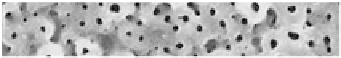
Oh
et al
. [63] showed that anodic oxidation of Ti at a very high
voltages result in a surface attractive for biomedical applications.
They kept anodization at a constant voltage of 180 V up to 60 min
in H
2
SO
4
+ H
3
PO
4
electrolytes. After anodizing at high voltages, a
porous titania surface is formed (Fig. 9.21) with a mixture of the
anatase and rutile phases on the Ti substrate (Fig. 9.22) [63].
Figure 9.21
SEM image of the surface morphologies of the anodic TiO
2
ilm prepared at 180 V in 0.9M H
2
SO
4
+ 0.1M H
3
PO
4
for 30
min [63].
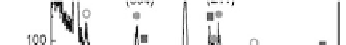
Figure 9.22
X-ray diffraction pattern of the anodic titania ilm formed at
180 V in 0.9M H
2
SO
4
+ 0.1M H
3
PO
4
for 30 min [63].