Biomedical Engineering Reference
In-Depth Information
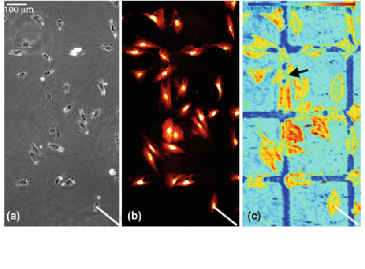
Fig. 7 Vascular smooth muscle cells grown on a substrate with 300 lm squares of fibronectin.
The cells had been fixed prior to imaging. a Phase-contrast micrograph. b Fluorescence
micrograph after Texas Red staining. c SPR-based micrograph (Adapted from [
38
])
as a suitable and in some respects even more powerful method for the visualization
and quantification of cell-substrate interactions.
Only recently the group of Peterson et al. [
38
,
39
] described an SPRM-based
analysis of remodeling processes of the ECM performed by vascular smooth muscle
cells during adherence, spreading and migration. By using a sophisticated optical
setup and a growth surface carrying fibronectin patterns, the group could simulta-
neously collect data about the cell density and distance from the matrix as well as
the amount of protein that was deposited or removed from the ECM, respectively.
Figure
7
c compares the SPRM image with conventional phase-contrast microscopy
(Fig.
7
a) and fluorescence microscopy (Fig.
7
b) of the same field of view.
While the sensitivity of the system is remarkably high (*20 ng/cm
2
), the lateral
resolution of the micrographs is still low (*2 lm) compared to other microscopic
techniques. Even though SPRM has not been used extensively to study cell-surface
adhesion, its unique technical features and readouts may drive further applications.
4.2 Mechanical Methods for Studying the Stability
of Cell-Surface Interactions
The mechanical stability of cell-surface interactions can be determined from
so-called detachment assays. In these assays, substrate-anchored cells are exposed
to mechanical forces that aim to detach the cells from the surface under study. The
Search WWH ::

Custom Search