Biomedical Engineering Reference
In-Depth Information
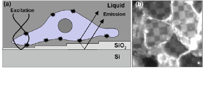
Fig. 4 a Schematic illustrating image formation in FLIC microscopy (adapted from [
24
]).
b Fluorescence micrograph of a cell grown on a FLIC substrate
the microscope either directly or after reflection at the silicon/silicon dioxide
interface. Since both the direct and the reflected fluorescent light are partly coherent,
interference occurs so that the intensity of the fluorescence emission is also
modulated by the optical path difference between membrane and silicon surface.
Taken together, the intensity of fluorophore excitation and the intensity of the
resulting fluorescence light are a function of the cell-substrate separation distance.
However, the relationship between the relative fluorescence intensity and the dis-
tance of the fluorophore to the silicon substrate surface is not unique but a damped
periodic function. The four different steps of silicon dioxide, serving as well-defined
spacers between the cell membrane and the reflecting silicon surface, are used to
provide four data pairs. The four different fluorescence intensities (cp. Fig.
4
b) are
analyzed using an optical theory, providing a distinct cell-substrate separation
distance with unprecedented precision of 1 nm [
24
,
25
]. However, FLIC is not a
label-free method and the cells may experience phototoxicity when repeated
experiments are performed to follow dynamic processes at the cell-surface junction.
4.1.3 Total Internal Reflection (Aqueous) Fluorescence Microscopy
Total internal reflection fluorescence microscopy (TIRF) [
26
] and total internal
reflection aqueous fluorescence microscopy (TIRAF) [
27
] are additional micro-
scopic techniques for visualizing the cell-surface junction of living cells as long as
they are grown on a transparent substrate. Both TIRF and TIRAF are subsumed
under the generic term evanescent field microscopy. In contrast to RICM
and similarly to FLIC, either the cell membrane (TIRF) or the incubation fluid
(TIRAF) requires fluorescent labeling. The cells under study are grown on a
transparent substrate that is illuminated from below with a laser beam. The laser
beam is aligned in such a way that it strikes the glass/liquid interface at an angle
bigger than or equal to the critical angle of total internal reflection h
crit
(Fig.
5
a).
Due to diffraction phenomena at the interface between an optically thicker and an
optically thinner medium, an evanescent electric field is generated at the surface
facing the liquid. Fluorophores attached to some component of the cell (TIRF) or
Search WWH ::

Custom Search