Biomedical Engineering Reference
In-Depth Information
(a)
(b)
1 mm
10 µm
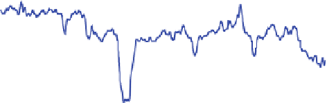
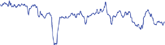
(c)
20
10
0
-10
-20
0
1
2
Length [mm]
Fig. 2 Surface structure of the carrier Sponceram HA
determined by a focus variation with an
InfiniteFocus microscope, b by scanning electron microscopy and c by profile measurements
From a macroscopic point of view, the surface of the Sponceram HA
received
exhibits a wavy structure with large local defects (Fig.
2
). Profile measurements
determined defects with a diameter of up to 150 lm and a depth of up to 100 lm.
The scanning electron microscopy (SEM) picture shows a disordered structure of
coarse grains exhibiting a diameter of about 1-2 lm and a length of 5-10 lm.
Density determination revealed a porous structure with relative density of 43%.
Relatively small macropores allow no ingrowth of cells; for bone tissue, for
example, pore diameters of more than 300 lm are recommended [
9
]. As cartilage
is an avascular tissue, a scaffold which supports angiogenesis by an interconnected
porosity with adequate pore diameters is not required for nutrient supply [
40
].
Using the cultivation principle described above, cartilage-carrier constructs
were generated on top of Sponceram HA
carrier and compared to cartilage
cultivated without a carrier. Significant differences could be observed in the
mass and the thickness of tissue-engineered cartilage. The wet weight of cartilage
grown without a carrier compared to cartilage grown on top of a carrier was
33.4 ± 1.7 mg and 18.6 ± 4.3 mg, respectively. The thickness was determined to
be 2.4 ± 0.1 mm for unsupported cartilage and 0.7 ± 0.2 mm for cartilage-carrier
constructs. With a factor of 3.5 for the Young's modulus and 2.3 for the glycos-
aminoglycan (GAG) content, values were significantly lower for the cartilage
grown on top of the carrier compared to cultivation without a carrier (Fig.
3
).


Search WWH ::

Custom Search