Biomedical Engineering Reference
In-Depth Information
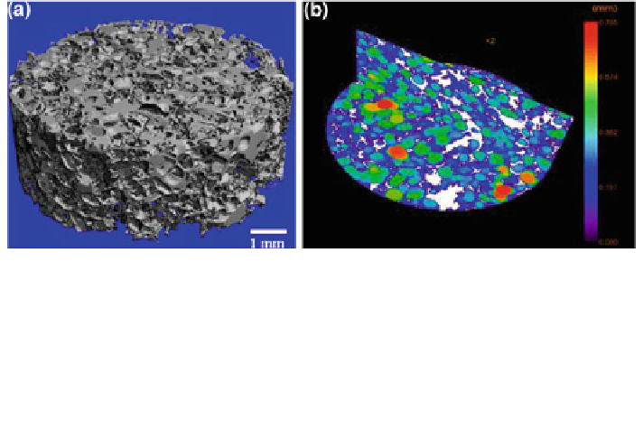
Fig. 8 a Micro-computed tomography image of a PDLLA scaffold fabricated by a sugar
template particulate leaching technique [
50
]. (Figure courtesy of R. Stämpfli, Empa St Gallen,
Switzerland). b Color-coded scaffold micro-architecture. The intermeshing, overlapping color
circles, i.e., color-coded pores (200-750 lm, 90
th
percentile: 600 lm), indicate high pore
interconnectivity and high porosity, which are both essential for cell penetration, proper
vascularization, bone tissue in-growth, waste removal, oxygen and nutrient delivery. The white
regions around the pores are PDLLA scaffold matrix areas. (Figure kindly provided by J.A. Sanz-
Herrera and I. Ochoa, University of Zaragoza, Spain)
are involved in angiogenesis. It has been shown, for example, that bioactive
glass stimulates the secretion of angiogenic growth factors in fibroblasts [
52
,
128
,
148
,
167
,
168
], the proliferation of endothelial cells [
50
,
53
], and the formation of
endothelial tubules [
53
].
In addition, in vivo results confirmed that BG is able to stimulate and promote
neo-vascularization [
49
,
103
-
105
,
169
]. For example, Leu et al. [
53
,
170
] filled
calvarial defects in Sprague-Dawley rats with 45S5 Bioglass
-impregnated
(1.2 mg) collagen sponges (volume = 0.05 cm
3
), using unloaded, empty sponges
as control. After 2 weeks of implantation, histological analyses of calvaria
demonstrated significantly greater neo-vascularization and vascular density within
defects treated with 45S5 BG (35 ± 16 vessels/mm
2
) than with collagen controls
alone (12 ± 2 vessels/mm
2
).
The angiogenic effect of bioactive glass has been shown to be much
more pronounced in bioactive glass-based scaffolds (i.e., loaded sponges [
170
],
discs [
171
], meshes [
169
], tubes [
172
], and porous glass-ceramic scaffolds
[
49
,
104
,
105
] than in composite structures incorporating and fully embedding
bioactive glass particles in polymer matrices (e.g., microsphere composites [
148
]
or foams [
52
,
173
]).
Day et al. [
52
], for example, found favorable angiogenic properties (i.e., greater
tissue infiltration and higher blood vessel formation) for compression-molded BG
composites compared to the corresponding unfilled polymer scaffolds. Interest-
ingly, the same authors found no difference in the number of blood vessels formed
in scaffolds prepared by thermally-induced phase separation technology. These
findings indicate that the geometry and morphology of the scaffold (pore orien-
tation, pore size, interconnectivity, strut thickness) can be used to control the
Search WWH ::

Custom Search