Biomedical Engineering Reference
In-Depth Information
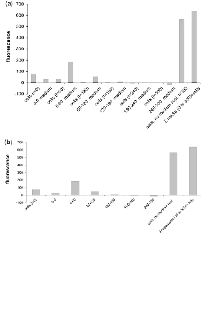
Fig. 4 Release of H
2
DCF and DCF after H
2
DCF-DA loading of A549 cells. A549 cells were
loaded with H
2
DCF-DA loading for 60 min. After two washes with HBSS medium, the
supernatant of some of the cultures was collected every 60 min and new HBSS was added.
Fluorescence of supernatants and cell cultures with medium replacement was measured every 60
min until 300 min after H
2
DCF-DA loading (485 nm extinction; 528 nm emission). DCF
fluorescence was seen in the supernatant and, at the start, also in the cells. The sum of the
fluorescence of all supernatants was similar to that of the cultures in which the medium was not
replaced. Thus it could be shown that DCF was formed in the cells and released in the
supernatant. After 300 min all media and cultures received H
2
O
2
(b). Fluorescence was measured
just before (t = 0) and 15 min and 18 h afterwards. Under the influence of H
2
O
2
, additional DCF
was formed in the supernatants, proving that H
2
DCF was also released by the cells. H
2
DCF-DA
was found to be insensitive to H
2
O
2
. Data are presented as mean ±SEM over three independent
experiments (A. Bruinink and U. Tobler, unpublished results)
Search WWH ::

Custom Search