Biomedical Engineering Reference
In-Depth Information
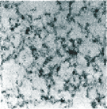
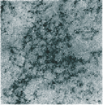
a
b
c
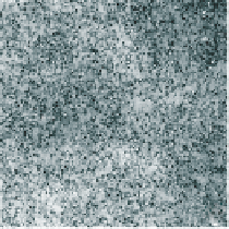
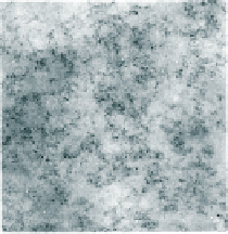
a
b
c
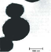
Figure 9.2
TEM images of BSA under various conditions: (a) pH 6.5, 10% w/w transparent gel in water;
(b) pH 6.5, 10% w/w turbid gel in 125 mN NaCl; (c) pH 5.1, 10% w/w coagulate at the isoelectric
point. The upper and lower images correspond to different magnifications, as indicated for (c).
Reproduced with permission from Clark et al.(
1981a
) © 1981 Munksgaard International
Publishers, a John Wiley & Sons subsidiary.
-
35
70 nm are created, although these structures are probably made up of rod-like
particles, also with widths ~3
5 nm (Carrotta et al.,
2001
). In agreement with this latter
picture, neutron scattering results at high ionic strength suggest the formation of spherical
structures ~20 nm in diameter (Aymard et al.,
1996b
).
Most recently of all, probe microscopy methods have been very widely employed
-
-
perhaps re
and atomic force
microscopy (AFM) has proved of great value. However, most of the latter work has
been applied to the class of
ecting the ready availability of such instrumentation
-
fibrillar gels now designated as amyloid gels (Hughes and
Dunstan,
2009
), discussed in more detail in
Section 9.5
.
Nevertheless, Gosal and co-workers (Gosal et al.,
2004a
) carried out an AFM analysis
of aggregates formed when
-Lg was heated for 24 h at 80°C at pH 7. The concentration
employed was 4% w/w, which is signi
β
cantly below the gel concentration. In agreement
with previous EM investigations, short rod-like particles were observed, with a typical
height of ~1.8 nm. However, this differs from previous EM estimates, where widths of
~3
5 nm were found (Clark et al.,
1981a
,
1981b
).
A great deal of work, especially including light scattering studies, has concentrated on
the speci
-
cs of the early aggregation stages. In some of these publications, a combination
of static and dynamic light scattering has been used to determine the sizes and shapes (i.e.
average molecular masses and radii of gyration) of the aggregates formed, including
fractal dimensions (Grif
n et al.,
1993
; Gimel et al.,
1994
;de
Kruif et al.,
1995
). From work of this kind it was concluded that the mode of aggregation
of
n and Grif
n,
1993
; Grif
β
-Lg at neutral pH and at a given ionic strength was essentially unaltered over a range

Search WWH ::

Custom Search