Biomedical Engineering Reference
In-Depth Information
Transverse
(edge-on crystals)
(
a
)
(
d
)
Channel
Hole
Overlap
D
40-50 A
°
Longitudinal (edge-on crystals)
(
b
)
Crystals face-on
Crystals edge-on
100 nm
(
e
)
M
M
C
(
c
)
5 nm
(
f
)
~67 nm
D
100 nm
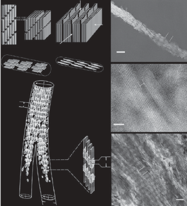
Figure 2.3
Structural organization of the mineralized collagen fi brils. (a) Model of
mineralized collagen fi brils showing the arrays of the plate-like mineral crystals
in the channels formed in staggered arranged collagen fi brils. (b) Face-on and
edge-on projections of the crystals in the mineralized fi bril. (c) The drawing of two
mineralized collagen fi brils in avian tendon. (d) TEM micrograph of an isolated
mineralized collagen fi bril from human dentin. (e) HRTEM micrograph of two
edge-on crystals in the mineralized collagen fi bril. (f) TEM micrograph of an array
of the mineralized fi brils from human dentin. Reprinted with permission from [34].
many methods including TEM, XRD, AFM, and Small angle X-ray scatter-
ing (SAXS) measurements have been used to analyze the HAp crystal size in
mineralized collagen fi brils, it is clear that the values of crystal size from dif-
ferent measurements are not consistent. TEM examination is considered to
be the most direct mean for visualizing these crystals. Robinson
et al.
, [22, 23]


Search WWH ::

Custom Search