Biomedical Engineering Reference
In-Depth Information
on the scaffolds [43]. GBMCs adhered, proliferated and showed osteogenic
differentiation on the scaffold. This was confi rmed after 14 days of culture
in osteogenic medium by alkaline phosphatase activity and immunohisto-
chemistry assays. The cells differentiated into chondrocytes after 21 days
of culture in chondrogenic medium. Their results indicated that bilayered
HA/CS scaffolds can be used for repair of osteochondral defects.
Zhang
et al.
cultured human fetal osteoblast cells (hFOB) on electros-
pun HA/CS nanocomposite scaffold. The cultured cells showed better cell
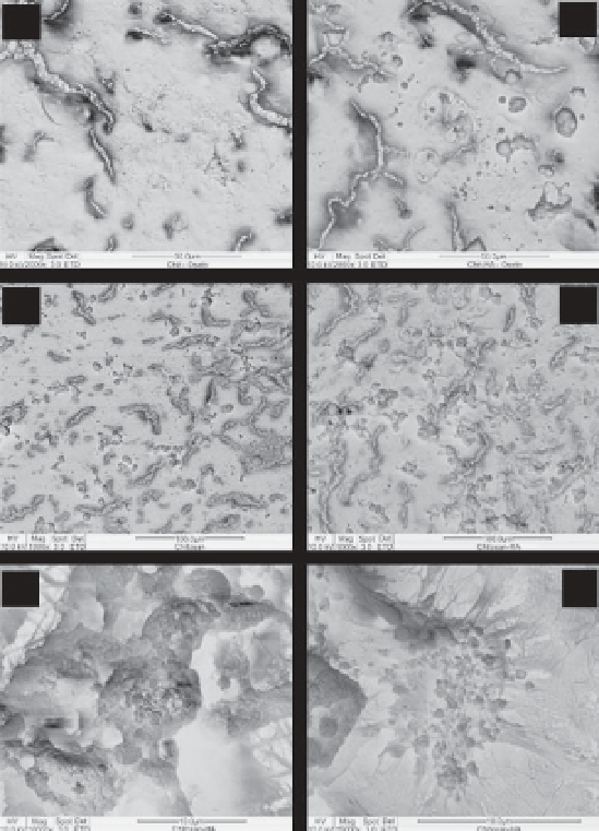
proliferation and mineral deposition than those cells cultured on plain chi-
tosan scaffolds as in Figure 14.6. This is attributed to the osteoconductive
(
b
)
(
a
)
(
c
)
(
d
)
(
e
)
(
f
)
Figure 14.6
Mineral depositions of hFOB on the electrospun nanofi brous
scaffolds: CTS, day 10 (a) and day 15 (c); HAp/CTS, day 10 (b) and day 15
(d); apatite-like morphology of deposit at higher magnifi cation (e); visible tiny
globular minerals and collagen bundles associated with a single hFOB cell
viewed at higher magnifi cation (f). Reprinted with permission from ref. [3].
Copyright (2008) Elsevier.
Search WWH ::

Custom Search